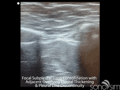
Bedside lung ultrasound may prove to be a sensitive early indicator of lung involvement in patients with coronavirus disease 2019 (COVID-19) infection. This greyscale ultrasound clip was taken from a COVID-19-positive patient on Day 1 of becoming symptomatic with fevers and myalgias. No cough was reported. The clip was taken along a transverse-oblique plane and demonstrates a focal hypoechoic focus consistent with a focal subpleural consolidation with adjacent overlying pleural thickening and pleural line discontinuity.
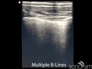
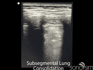
While there is still more research to be done, several recent studies have documented characteristic computed tomography (CT) and ultrasound findings in COVID-19 patients. The finding of peripheral, subpleural consolidations may prove to be an early indicator of pulmonary involvement, which may aid in the initial diagnosis, triaging, or disposition of patients presenting within the first several days of symptom onset (Chung et al.). Additional non-specific lung ultrasound findings that have been described in COVID-19 patients include (1) pleural thickening and irregularity, (2) focal and diffuse B-lines, (3) consolidations, and (4) pleural effusions (Huang et al., Peng et al.). Ultrasound seems to be an effective way of monitoring disease progression.

Published CT lung findings in COVID-19 patients are variable but often included one or more of the following characteristics: ground-glass opacities, consolidative pulmonary opacities, opacities with rounded morphology, and peripheral lung distribution (Chung et al., Wang et al.). Pleural effusions, cavitary lesions, and mediastinal adenopathy were less common findings (Chung et al.).
Given the evolving understanding of the role that aerosol and droplet spread play in severe and critical cases that are acute respiratory syndrome coronavirus 2 (SARS-COV-2) transmission, the degree and method for the ultrasound machine and probe disinfection must be robust. Disinfection protocols, use of protective transducer covers, and day-to-day execution will depend upon available resources, institutional guidelines, original equipment manufacturer (OEM) recommendations, and federal guidelines.
Key Points
- Focal subpleural consolidation with adjacent overlying pleural thickening and pleural line discontinuity may be an early finding with COVID-19 infection (Chung et al.)
- Additional non-specific lung ultrasound findings in patients with COVID-19 infections include pleural thickening and irregularity, focal and diffuse B-lines, consolidations, and pleural effusions (Huang et al., Peng et al.)
- Disinfection protocols, use of protective transducer covers, and day-to-day execution will depend upon available resources, institutional guidelines, OEM recommendations, and federal guidelines.
Chung M, Bernheim A, Mei X, et al. CT imaging features of 2019 novel Coronavirus (2019-nCoV). Radiology. 2020 Apr;295(1):202-207. doi: 10.1148/radiol.2020200230. Epub 2020 Feb 4.
Huang Y, Wang S, Liu Y, et al. A preliminary study on the ultrasonic manifestations of peripulmonary lesions of non-critical novel coronavirus pneumonia (COVID-19). [Prepint]. 2020 [cited 2020 Mar 23]: [14 p.]. Available from: https://ssrn.com/abstract=3544750 or http://dx.doi.org/10.2139/ssrn.3544750
Peng, QY, Wang XT, Zhang LN; Chinese Critical Care Ultrasound Study Group. Findings of lung ultrasonography of novel coronavirus pneumonia during the 2019–2020 epidemic. Intensive Care Medicine. 2020 Mar: 1-2. doi: 10.1007/s00134-020-05996-6. [Epub ahead of print]
Wang YXJ, Liu WH, Yang M, et al. The role of CT for Covid-19 patient management remains poorly defined. Ann Transl Med. 2020 Feb;8(4):145. doi:10.21037/atm.2020.02.71
*Information from this website is for informational and learning purposes. It is not a substitute for professional medical advice, diagnosis, or treatment, but is intended to share real-time case studies and academic articles within the medical education community.