


-
Anatomy & Physiology
-
Core Clinical
-
Advanced Clinical
-
U/S-Guided Procedures
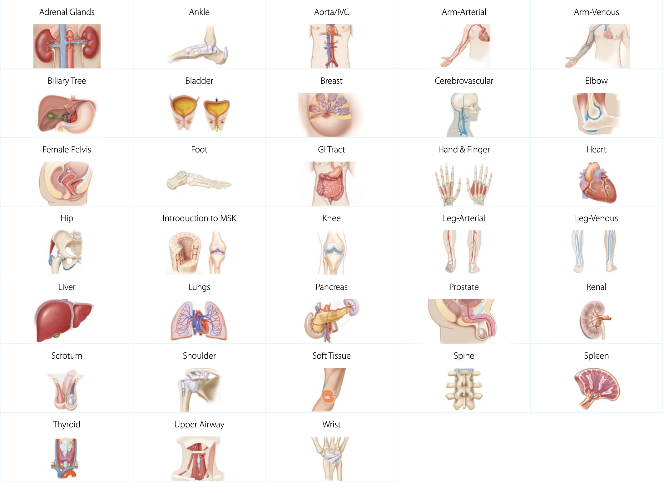
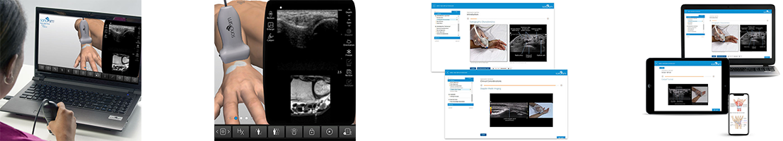
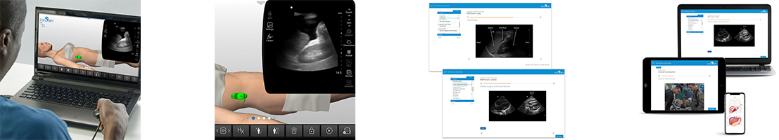
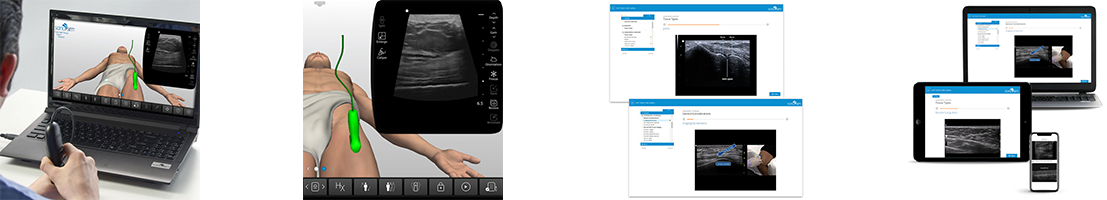
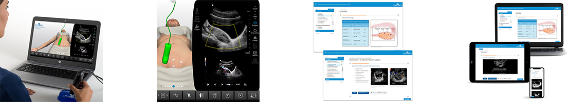
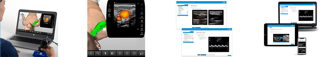
Anatomy & Physiology Ultrasound Training
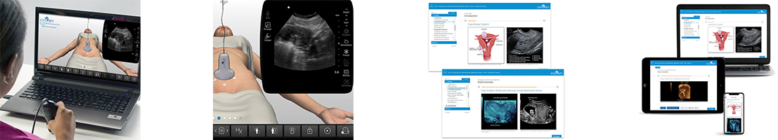
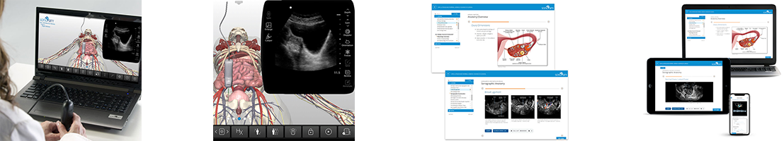
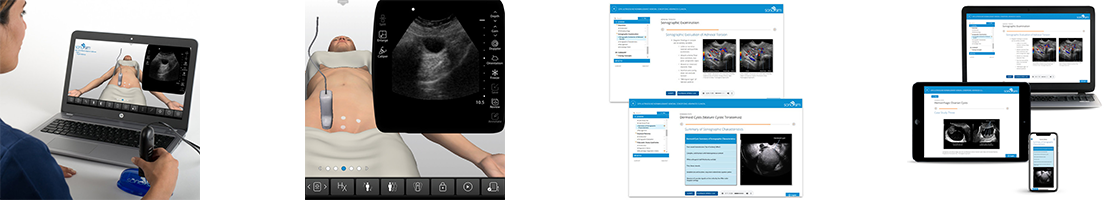
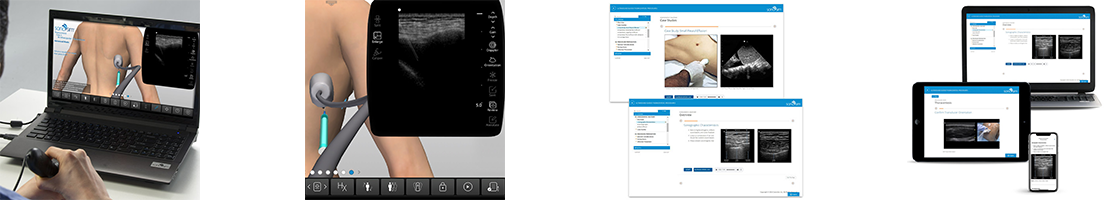
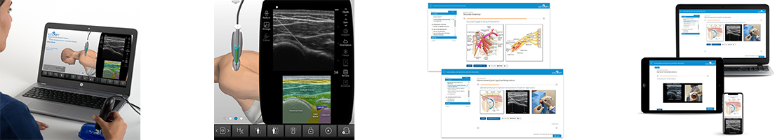
Our A&P ultrasound training provides a strong foundation of ultrasound knowledge, specific to anatomical regions, organs, and structures. Our expert-led ultrasound courses cover everything from regional anatomy & physiology to sonographic anatomy, scanning technique, and imaging tips & pitfalls. Relevant scanning cases from real patients are accessible in the SonoSimulator for each topic.
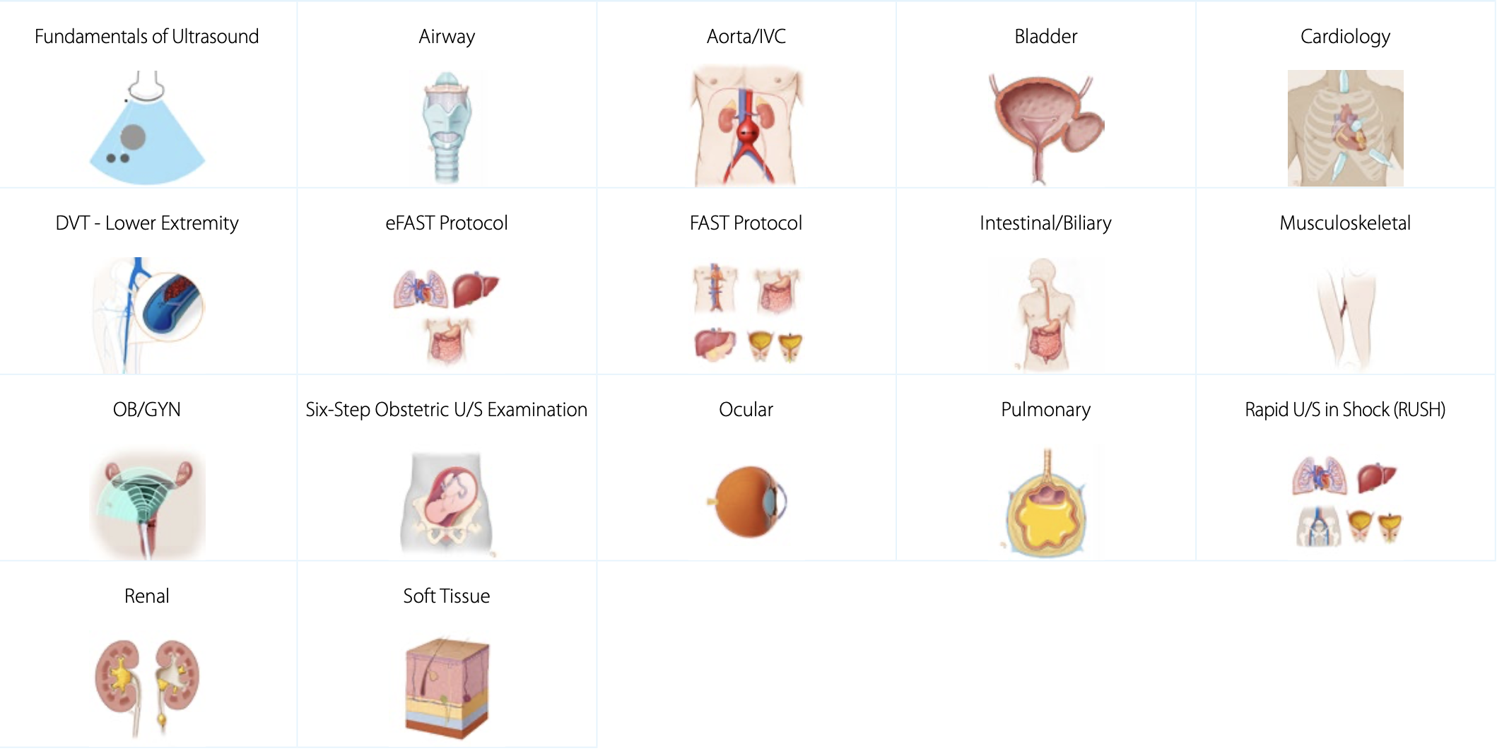
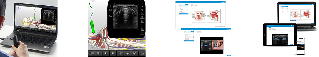
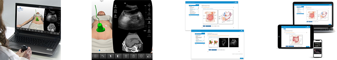
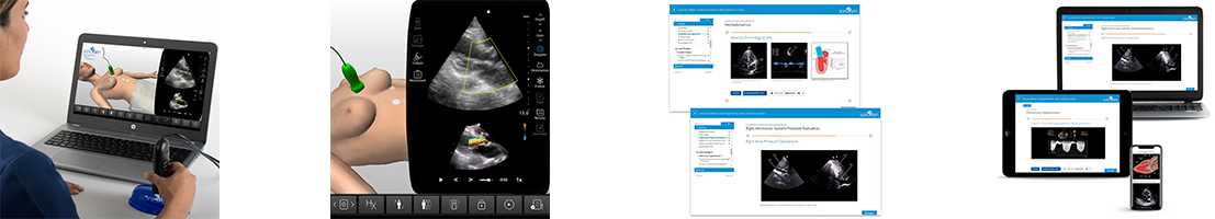
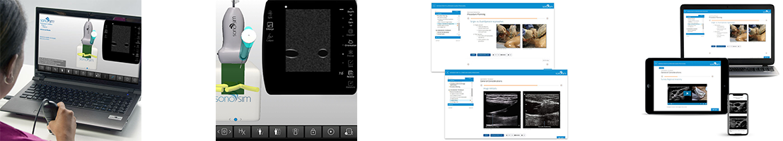
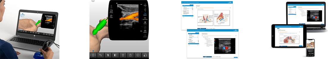
Core Clinical Ultrasound Training
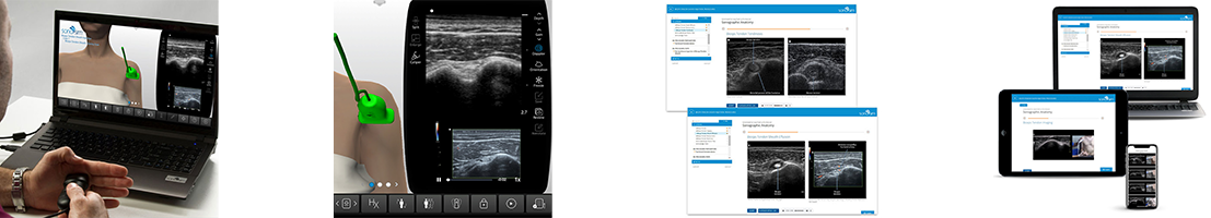
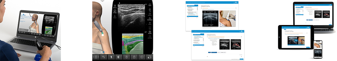
Our clinical ultrasound training topics provide in-depth comprehension of the necessary components to accurately assess and diagnose using ultrasound. We cover exam indications, regional anatomy, sonographic anatomy, and sonographic technique. Our ultrasound courses use pathologic case studies and imaging tips & pitfalls to give you an in-depth understanding of how diseases present in ultrasound imaging. Associated scanning cases from real patients are included and accessible in the SonoSimulator. This topic category supports both diagnostic medical sonography (DMS) learners as well as medical practitioners looking for point-of-care ultrasound (POCUS) training.
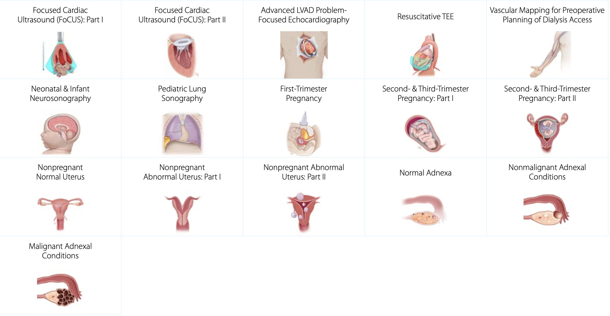
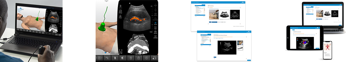
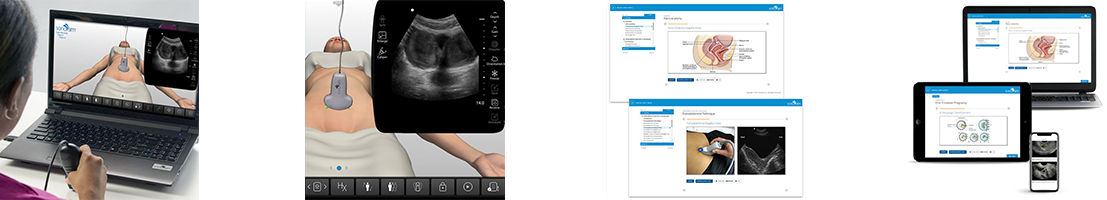
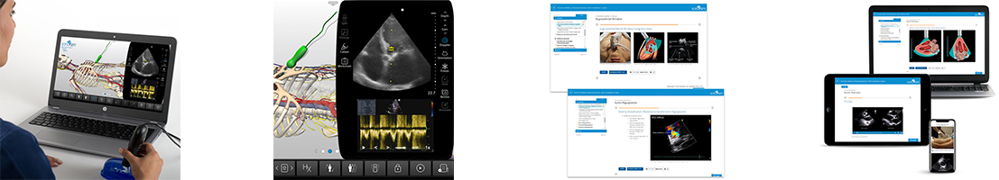
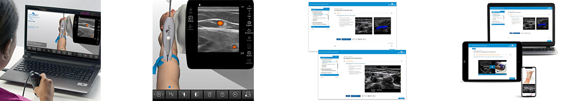
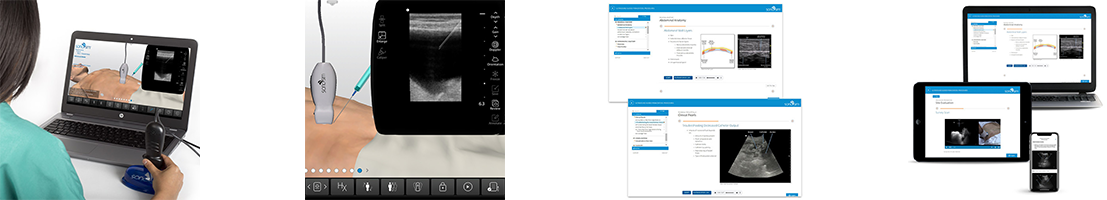
Advanced Clinical Ultrasound Training
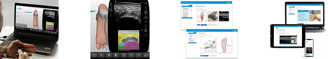
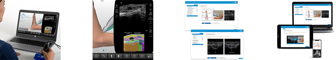
Our advanced clinical ultrasound topics cover complex diagnoses and sonographic applications. This is an ever-expanding list of topics with a focus on specific pathologic conditions, providing learners a deep understanding of ultrasound techniques for assessing a variety of complex medical conditions. Each topic is accompanied with 10-20 scanning cases from real patients, that are accessed in the SonoSimulator.
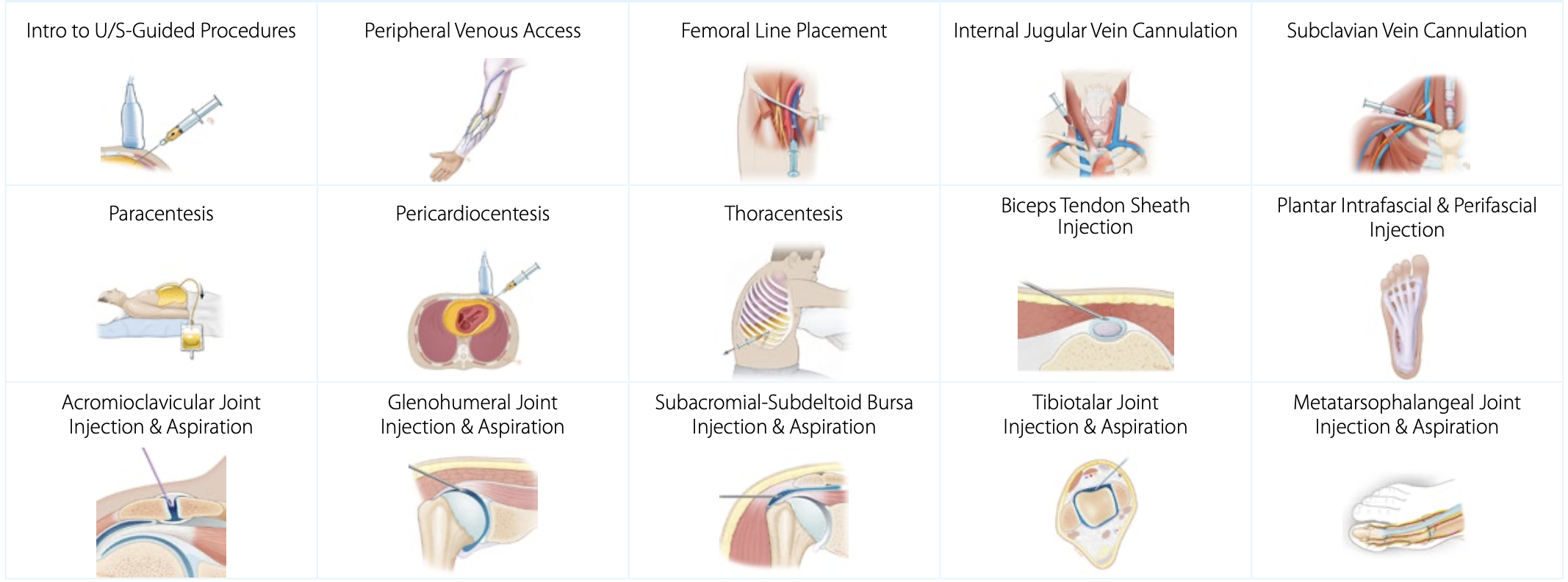
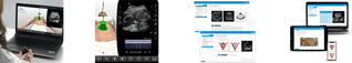
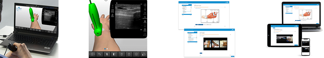
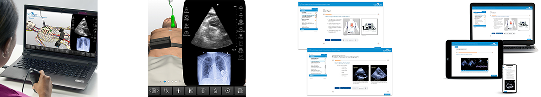
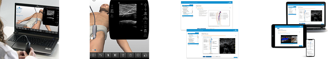
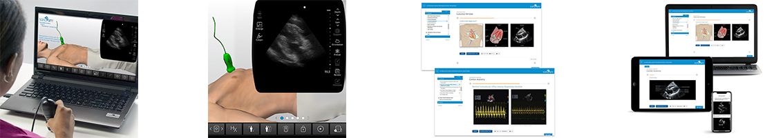
Ultrasound-Guided Procedure Training
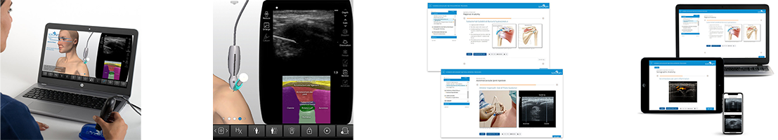
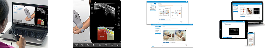
Ultrasound-guided procedures are increasingly used to minimize complications and improve patient care. SonoSim ultrasound procedure training covers patient positioning, procedural steps, imaging adjuncts, potential complications, and more. Scanning cases from real patients are also provided with these topics, to support unlimited practice of the cognitive tasks required to perform ultrasound-guided procedures. Practice as much as you like, risk-free - anytime, anywhere.


Adrenal Glands Anatomy & Physiology Ultrasound Training
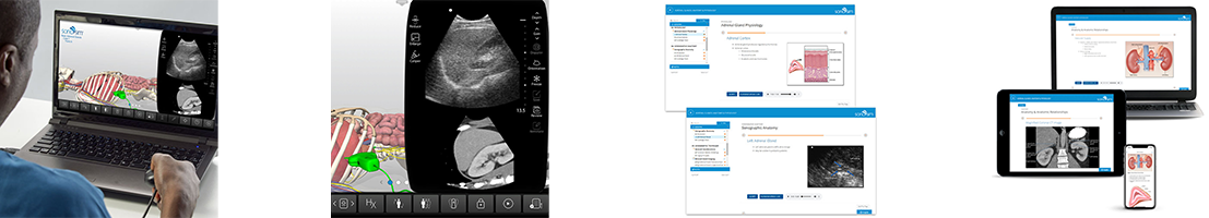
Learn the relationship of traditional illustrative and sonographic anatomy of the adrenal glands and how to interpret ultrasound imaging of the region. Learn sonographic techniques and scanning approaches used in ultrasound imaging to assess the adrenals. Learn to identify and characterize the adrenal glands with ultrasonography.
Course Highlights |
SonoSimulator Scanning Cases |
|
The course in this module covers topics including:
|
The 3 real patient scanning cases in this module include and cover:
|

Ankle Anatomy & Physiology Ultrasound Training
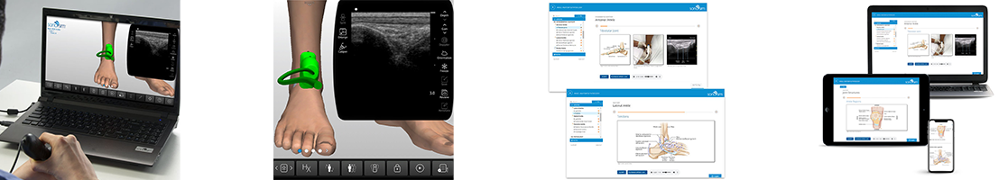
Learn the relationship of traditional illustrative and sonographic anatomy of the ankle and how to interpret ultrasound imaging of the ankle. Learn sonographic techniques and scanning approaches used in ultrasound imaging to assess the ankle. Learn to identify and characterize the ankle with ultrasonography.
Course Highlights |
SonoSimulator Scanning Cases |
|
The course in this module covers topics including:
|
The 4 real patient scanning cases in this module include and cover:
|

Aorta & Inferior Vena Cava ( IVC ) Anatomy & Physiology Ultrasound Training
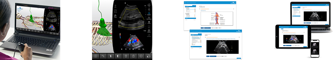
Learn the relationship of traditional illustrative and sonographic anatomy of the aorta and inferior vena cava (IVC) and how to interpret ultrasound imaging of the aorta & IVC. Learn sonographic techniques and scanning approaches used in ultrasound imaging to assess the aorta and inferior vena cava. Learn to identify and characterize the aorta / IVC with ultrasonography.
Course Highlights |
SonoSimulator Scanning Cases |
|
The course in this module covers topics including:
|
The 5 real patient scanning cases in this module include and cover:
|
Arm Arterial Anatomy & Physiology Ultrasound Training
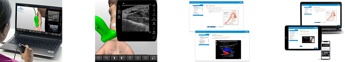
Learn the relationship of traditional illustrative and sonographic anatomy of the arm and how to interpret ultrasound imaging of the upper extremity arterial system. Learn sonographic techniques and scanning approaches used in ultrasound imaging of the upper extremity arterial system. Learn to identify and characterize the macroscopic and regional anatomical components of the upper extremity arterial system with ultrasonography.
Course Highlights |
SonoSimulator Scanning Cases |
|
The course in this module covers topics including:
|
The 5 real patient scanning cases in this module include and cover:
|
Arm Venous Anatomy & Physiology Ultrasound Training
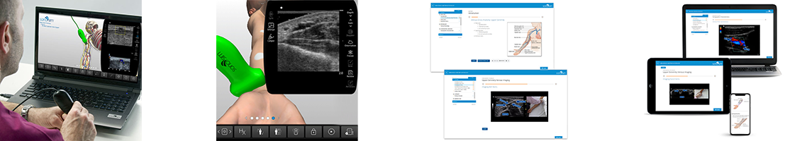
Learn the relationship of traditional illustrative and sonographic anatomy of the arm and how to interpret ultrasound imaging of arm veins. Learn sonographic techniques and scanning approaches used in ultrasound imaging to assess the venous system of the upper extremities. Learn to identify and characterize arm veins with ultrasonography.
Course Highlights |
SonoSimulator Scanning Cases |
|
The course in this module covers topics including:
|
The 5 real patient scanning cases in this module include and cover:
|
Biliary Tree Anatomy & Physiology Ultrasound Training
Learn the relationship of traditional illustrative and sonographic anatomy of the biliary tree and how to interpret ultrasound imaging of the gallbladder, vascular supply and surrounding structures. Learn sonographic techniques and scanning approaches used in ultrasound imaging to assess the biliary tree including bile storage and enterohepatic circulation. Learn to identify and characterize the macroscopic and regional anatomical components of the biliary tract with ultrasonography.
Course Highlights |
SonoSimulator Scanning Cases |
The course in this module covers topics including:
|
The 3 real patient scanning cases in this module include and cover:
|
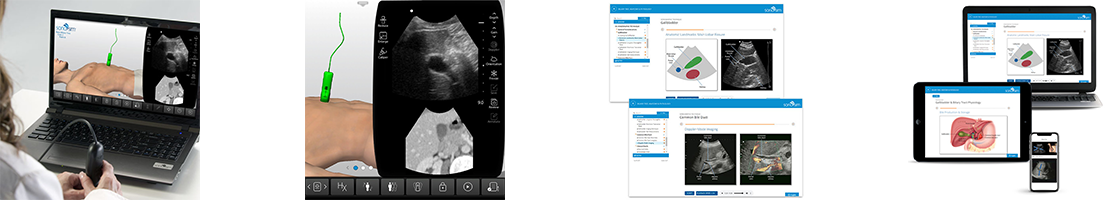
Bladder Anatomy & Physiology Ultrasound Training
Learn the relationship of traditional illustrative and sonographic anatomy of the bladder and how to interpret ultrasound imaging of the bladder and surrounding structures. Learn sonographic techniques and scanning approaches used in ultrasound imaging to assess the bladder including bladder volume measurements. Learn to identify and characterize the bladder with ultrasonography.
Course Highlights |
SonoSimulator Scanning Cases |
|
The course in this module covers topics including:
|
The 5 real patient scanning cases in this module include and cover:
|
Breast Anatomy & Physiology Ultrasound Training
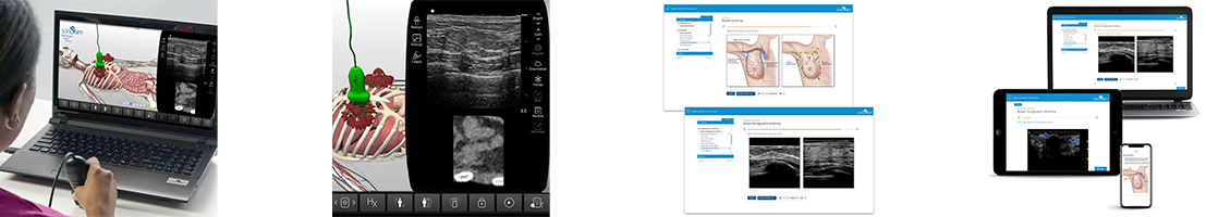
Learn the relationship of traditional illustrative and sonographic anatomy and how to interpret ultrasound imaging of the breast. Learn sonographic techniques and scanning approaches used in ultrasound imaging to assess the breast including breast layers, normal anatomical variants and age-related changes. Learn to identify and characterize the breast with ultrasonography.
Course Highlights |
SonoSimulator Scanning Cases |
|
The course in this module covers topics including:
|
The 3 real patient scanning cases in this module include and cover:
|
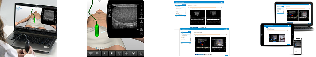
Cerebrovascular Anatomy & Physiology Ultrasound Training
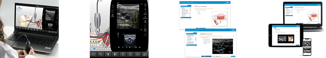
Learn the relationship of traditional illustrative and sonographic anatomy of the cerebral vasculature and how to interpret cerebrovascular ultrasound imaging. Learn sonographic techniques and scanning approaches used in ultrasound imaging to assess cerebral vasculature. Learn to identify and characterize cerebral vasculature with ultrasonography.
Course Highlights |
SonoSimulator Scanning Cases |
|
The course in this module covers topics including:
|
The 5 real patient scanning cases in this module include and cover:
|
Elbow Anatomy & Physiology Ultrasound Training
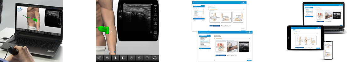
Learn the relationship of traditional illustrative and sonographic anatomy and how to interpret ultrasound imaging of the elbow. Learn sonographic techniques and scanning approaches used in ultrasound imaging to assess the elbow. Learn to identify and characterize the elbow with ultrasonography.
Course Highlights |
SonoSimulator Scanning Cases |
|
The course in this module covers topics including:
|
The 4 real patient scanning cases in this module include and cover:
|
Female Pelvis Anatomy & Physiology Ultrasound Training
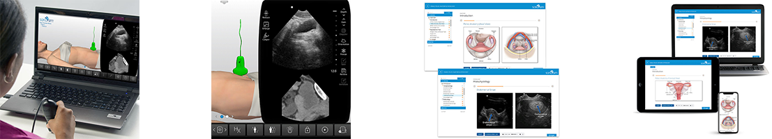
Learn the relationship of traditional illustrative and sonographic anatomy and how to interpret ultrasound imaging of the female pelvis. Learn sonographic techniques and scanning approaches used in ultrasound imaging to assess the female pelvis. Learn to identify and characterize the female pelvis with ultrasonography.
Course Highlights |
SonoSimulator Scanning Cases |
|
The course in this module covers topics including:
|
The 3 real patient scanning cases in this module include and cover:
|
Foot Anatomy & Physiology Ultrasound Training
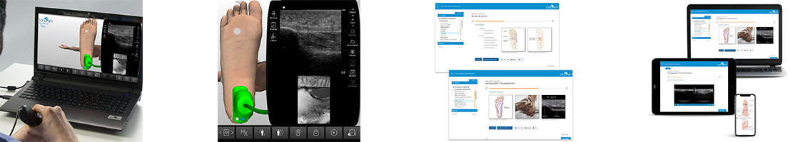
Learn the relationship of traditional illustrative and sonographic anatomy and how to interpret ultrasound imaging of the foot. Learn sonographic techniques and scanning approaches used in ultrasound imaging to assess the foot. Learn to identify and characterize the foot with ultrasonography.
Course Highlights |
SonoSimulator Scanning Cases |
|
The course in this module covers topics including:
|
The 3 real patient scanning cases in this module include and cover:
|
GI Tract Anatomy & Physiology Ultrasound Training
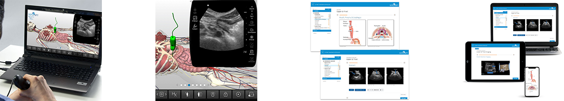
Learn the relationship of traditional illustrative and sonographic anatomy and how to interpret ultrasound imaging of the gastrointestinal tract, including the mouth, pharynx, esophagus, stomach, intestines, and colon. Learn sonographic techniques and scanning approaches used in ultrasound imaging to assess the GI tract. Learn to identify and characterize the gastrointestinal system using ultrasonography.
Course Highlights |
SonoSimulator Scanning Cases |
|
The course in this module covers topics including:
|
The 3 real patient scanning cases in this module include and cover:
|
Hand & Finger Anatomy & Physiology Ultrasound Training
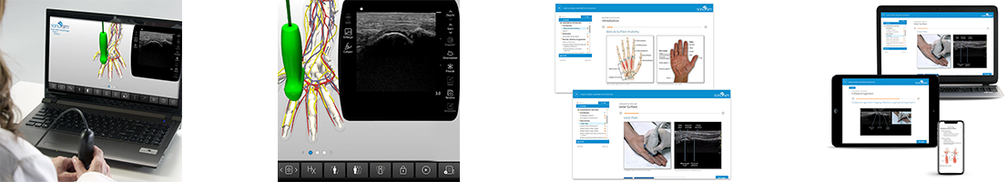
Learn the relationship of traditional illustrative and sonographic anatomy of the hand and fingers and how to interpret ultrasound imaging of the hand and fingers. Learn sonographic techniques and scanning approaches used in ultrasound imaging to assess the hand and fingers. Learn to identify and characterize the hand and fingers with ultrasonography.
Course Highlights |
SonoSimulator Scanning Cases |
|
The course in this module covers topics including:
|
The 5 real patient scanning cases in this module include and cover:
|
Heart Anatomy & Physiology Ultrasound Training
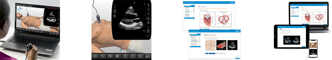
Learn the relationship of traditional illustrative and sonographic anatomy and how to interpret ultrasound imaging of the heart. Learn sonographic techniques and scanning approaches used in cardiac ultrasound imaging. Learn characterize the heart using ultrasonography.
Course Highlights |
SonoSimulator Scanning Cases |
|
The course in this module covers topics including:
|
The 5 real patient scanning cases in this module include and cover:
|
Hip Anatomy & Physiology Ultrasound Training
Learn the relationship of traditional illustrative and sonographic anatomy of the hip and how to interpret ultrasound imaging of the hip. Learn sonographic techniques and scanning approaches used in ultrasound imaging to assess the hip. Learn to identify and characterize the hip with ultrasonography.
Course Highlights |
SonoSimulator Scanning Cases |
|
The course in this module covers topics including:
|
The 4 real patient scanning cases in this module include and cover:
|
Knee Anatomy & Physiology Ultrasound Training
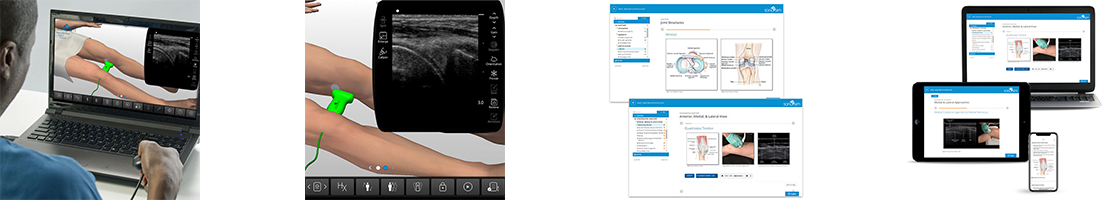
Learn how to use ultrasound in the evaluation of the knee, including knee anatomy and physiology, and sonographic anatomy of the knee. Optimal transducer selection, patient positioning, imaging approaches, and techniques for sonographically evaluating the knee are discussed.
Course Highlights |
SonoSimulator Scanning Cases |
|
The course in this module covers topics including:
|
The 4 real patient scanning cases in this module include and cover:
|
Leg Arterial Anatomy & Physiology Ultrasound Training
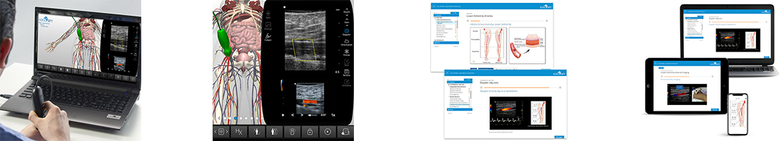
Learn how to use ultrasound in the evaluation of the lower extremity arterial system, including lower extremity arterial anatomy and physiology and sonographic anatomy of the lower extremity arterial system. Optimal transducer selection, patient positioning, imaging approaches, and techniques for sonographically evaluating the lower extremity arterial system are discussed.
Course Highlights |
SonoSimulator Scanning Cases |
|
The course in this module covers topics including:
|
The course in this module covers topics including:
|
Leg Venous Anatomy & Physiology Ultrasound Training
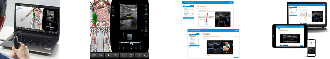
Learn how to use ultrasound in the evaluation of the lower extremity vasculature, including lower extremity venous anatomy and physiology, and sonographic anatomy of the lower extremity venous system. Optimal transducer selection, patient positioning, imaging approaches, and techniques for sonographically evaluating the lower extremity venous system are discussed.
Course Highlights |
SonoSimulator Scanning Cases |
|
The course in this module covers topics including:
|
The 5 real patient scanning cases in this module include and cover:
|
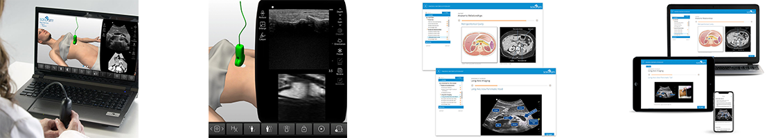
Liver Anatomy & Physiology Ultrasound Training
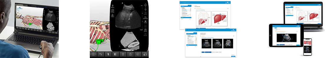
Learn how to use ultrasound in the evaluation of the liver, including anatomy and physiology, and sonographic anatomy of the liver. Optimal transducer selection, patient positioning, imaging approaches, and techniques for sonographically evaluating the liver are discussed.
Course Highlights |
SonoSimulator Scanning Cases |
|
The course in this module covers topics including:
|
The 3 real patient scanning cases in this module include and cover:
|
Lungs Anatomy & Physiology Ultrasound Training
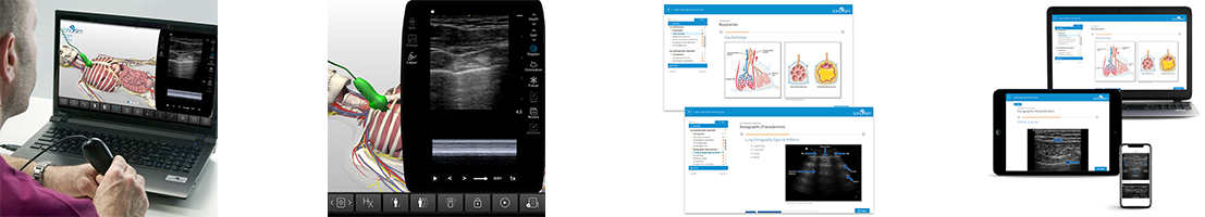
Learn how to use ultrasound in the evaluation of the lungs, including lung anatomy and physiology, and sonographic anatomy of the lungs. Optimal transducer selection, patient positioning, imaging approaches, and techniques for sonographically evaluating the lungs are discussed.
Course Highlights |
SonoSimulator Scanning Cases |
|
The course in this module covers topics including:
|
The 5 real patient scanning cases in this module include and cover:
|
MSK Anatomy & Physiology Introductory Ultrasound Training
Learn the relationship of traditional illustrative and sonographic anatomy of the musculoskeletal system, including how to interpret ultrasound imaging of the musculoskeletal system. Learn sonographic techniques and scanning approaches used in ultrasound imaging to assess the musculoskeletal system. Learn to identify and characterize the components of the musculoskeletal system with ultrasonography.
Course Highlights |
SonoSimulator Scanning Cases |
|
The course in this module covers topics including:
|
The 4 real patient scanning cases in this module include and cover:
|
Pancreas Anatomy & Physiology Ultrasound Training
Learn how to use ultrasound to evaluate the pancreas, including pancreas anatomy and physiology and sonographic anatomy of the pancreas. Optimal transducer selection, patient positioning, imaging approaches, and techniques for sonographically evaluating the pancreas are discussed.
Course Highlights |
SonoSimulator Scanning Cases |
|
The course in this module covers topics including:
|
The 3 real patient scanning cases in this module include and cover:
|
Prostate Anatomy & Physiology Ultrasound Training
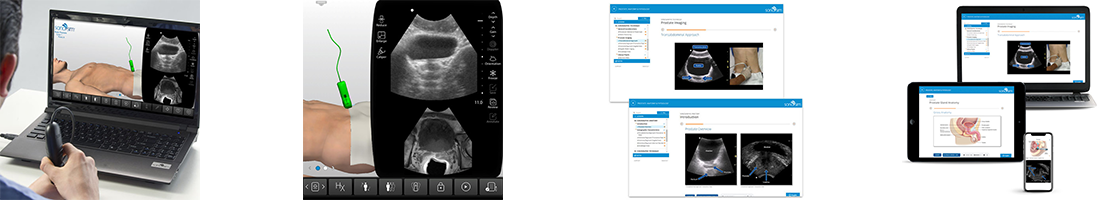
Learn how to use ultrasound in the evaluation of the prostate, including prostate anatomy and physiology, and sonographic anatomy of the prostate. Optimal transducer selection, patient positioning, imaging approaches, and techniques for sonographically evaluating the prostate are discussed.
Course Highlights |
SonoSimulator Scanning Cases |
|
The course in this module covers topics including:
|
The 3 real patient scanning cases in this module include and cover:
|
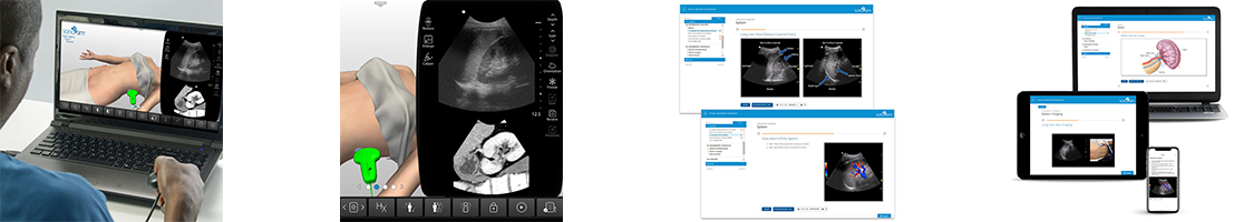
Renal Anatomy & Physiology Ultrasound Training
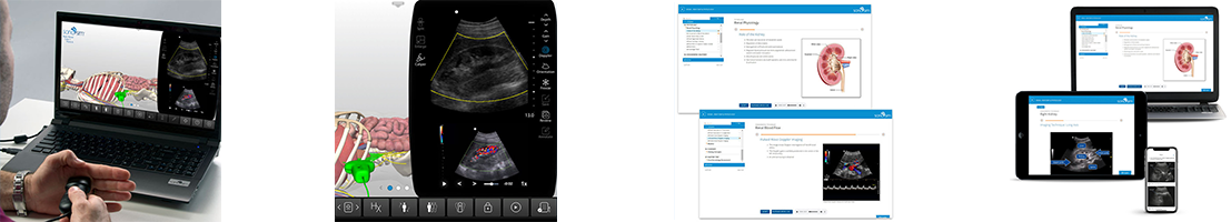
Learn how to use ultrasound in the evaluation of the kidneys, including kidney anatomy and physiology and sonographic anatomy of the kidneys. Optimal transducer selection, patient positioning, imaging approaches, and techniques for sonographically evaluating the kidneys are discussed.
Course Highlights |
SonoSimulator Scanning Cases |
|
The course in this module covers topics including:
|
The 5 real patient scanning cases in this module include and cover:
|
Scrotum Anatomy & Physiology Ultrasound Training
Learn how to use ultrasound in the evaluation of the scrotum, including scrotum anatomy and physiology and sonographic anatomy of the scrotum. Optimal transducer selection, patient positioning, imaging approaches, and techniques for sonographically evaluating the scrotum are discussed.
Course Highlights |
SonoSimulator Scanning Cases |
|
The course in this module covers topics including:
|
The 3 real patient scanning cases in this module include and cover:
|
Shoulder Anatomy & Physiology Ultrasound Training
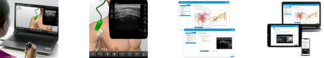
Learn to use ultrasound in the evaluation of the shoulder, including shoulder anatomy and physiology, and sonographic anatomy of the shoulder. Optimal transducer selection, patient positioning, imaging approaches, and techniques for sonographically evaluating the shoulder are discussed.
Course Highlights |
SonoSimulator Scanning Cases |
|
The course in this module covers topics including:
|
The 3 real patient scanning cases in this module include and cover:
|
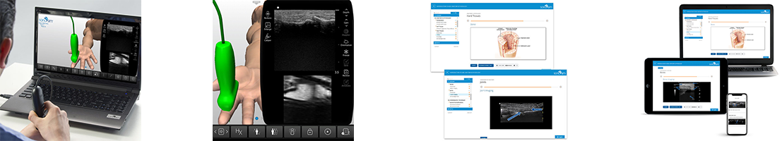
Soft Tissue Anatomy & Physiology Ultrasound Training
Learn how to use ultrasound in the evaluation of the soft tissues, including soft tissue anatomy and physiology and sonographic anatomy of soft tissues. Optimal transducer selection, patient positioning, imaging approaches, and techniques for sonographically evaluating soft tissue are discussed.
Course Highlights |
SonoSimulator Scanning Cases |
|
The course in this module covers topics including:
|
The 4 real patient scanning cases in this module include and cover:
|
Spine Anatomy & Physiology Ultrasound Training
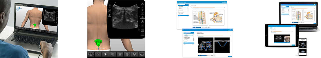
Learn how to use ultrasound in the evaluation of the spine, including spine anatomy and physiology, and sonographic anatomy of the spine. Optimal transducer selection, patient positioning, imaging approaches, and techniques for sonographically evaluating the spine are discussed.
Course Highlights |
SonoSimulator Scanning Cases |
|
The course in this module covers topics including:
|
The 3 real patient scanning cases in this module include and cover:
|
Spleen Anatomy & Physiology Ultrasound Training
Learn how to use ultrasound in the evaluation of the spleen, including spleen anatomy and physiology, and sonographic anatomy of the spleen. Optimal transducer selection, patient positioning, imaging approaches, and techniques for sonographically evaluating the spleen are discussed.
Course Highlights |
SonoSimulator Scanning Cases |
|
The course in this module covers topics including:
|
The 3 real patient scanning cases in this module include and cover:
|
Thyroid Anatomy & Physiology Ultrasound Training
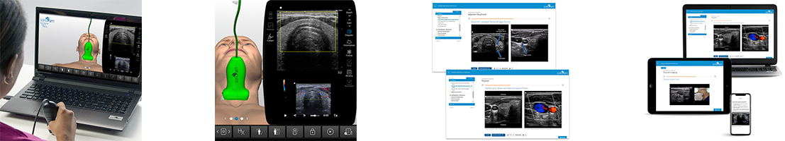
Learn how to use ultrasound in the evaluation of the thyroid, including thyroid anatomy and physiology, and sonographic anatomy of the thyroid. Optimal transducer selection, patient positioning, imaging approaches, and techniques for sonographically evaluating the thyroid are discussed.
Course Highlights |
SonoSimulator Scanning Cases |
|
The course in this module covers topics including:
|
The 5 real patient scanning cases in this module include and cover:
|
Upper Airway Anatomy & Physiology Ultrasound Training
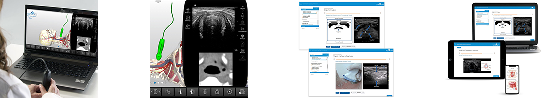
Learn how to use ultrasound in the evaluation of the upper airway, including upper airway anatomy and physiology, and sonographic anatomy of the upper airway. Optimal transducer selection, patient positioning, imaging approaches, and techniques for sonographically evaluating the upper airway are discussed.
Course Highlights |
SonoSimulator Scanning Cases |
|
The course in this module covers topics including:
|
The 5 real patient scanning cases in this module include and cover:
|
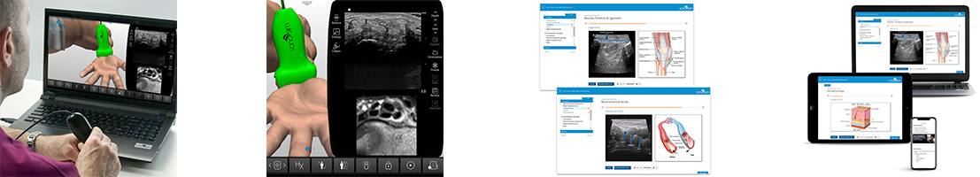
Wrist Anatomy & Physiology Ultrasound Training
Learn how to use ultrasound in the evaluation of the wrist, including wrist anatomy and physiology, and sonographic anatomy of the wrist. Optimal transducer selection, patient positioning, imaging approaches, and techniques for sonographically evaluating the wrist are discussed.
Course Highlights |
SonoSimulator Scanning Cases |
|
The course in this module covers topics including:
|
The SonoSimulator® helps develop and maintain the critical visuomotor and visuospatial skills that are central to image acquisition and interpretation with real patient imagery, expert tutorials on-demand, and real-time feedback on success. The 3 real patient scanning cases in this module include and cover:
|


Airway Clinical Ultrasound Training
Learn the basic principles and physics of point-of-care airway ultrasound imaging and how to optimize image acquisition of the human airway. Learn to evaluate the airway in an emergency and direct mainstem intubation. Learn how to use ultrasound in the evaluation of the emergent airway to confirm tracheal intubation.
Course Highlights |
SonoSimulator Scanning Cases |
|
The course in this module covers topics including:
|
The 10 real patient scanning cases in this module include and cover:
|
Aorta & Inferior Vena Cava ( IVC ) Clinical Ultrasound Training
Learn how to evaluate aortic anatomy and identify abdominal aortic aneurysms (AAAs) with ultrasound. How to evaluate IVC anatomy and how to estimate central venous pressure (CVP) are also discussed. Practical application factors including transducer selection, imaging techniques, and scanning approaches are covered.
Course Highlights |
SonoSimulator Scanning Cases |
|
The course in this module covers topics including:
|
The 20 real patient scanning cases in this module include and cover:
|
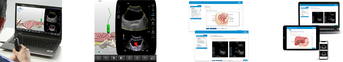
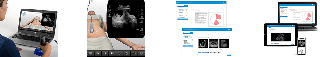
Basic Obstetric Ultrasound Exam - Six-Step Approach
This POCUS training module teaches a six-step approach for assessing pregnancy at the point of care. In six-steps, learn how to use ultrasound to identify conditions in the second and third trimester of pregnancy that have immediate patient care implications and/or that require planning for subsequent perinatal care and delivery in a facility that is equipped and staffed to deal with obstetric complications. The basic principles for assessing fetal presentation and lie, fetal cardiac activity, presence of multiple pregnancy, placental location within the uterus, amniotic fluid volume, and fetal biometry are discussed in detail. Designed for point-of-care healthcare professionals that care for pregnant patients. This module is also of particular relevance for maternal health workers practicing in resource-constrained environments.
Course Highlights |
SonoSimulator Scanning Cases |
|
The course in this module covers topics including:
|
The 10 real patient scanning cases in this module include and cover:
|
Bladder Clinical Ultrasound Training
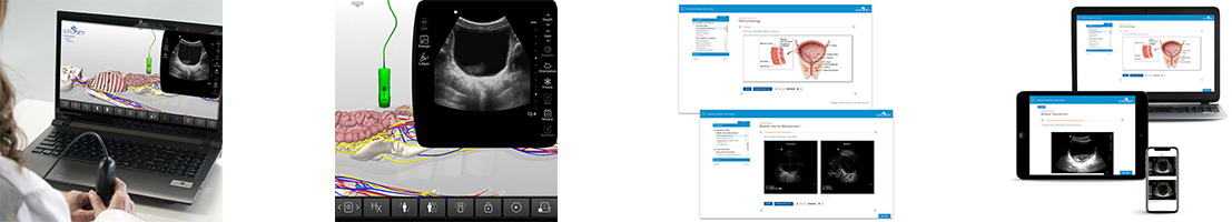
Learn how to determine bladder volume measurements, including post-void residual volume, with ultrasound. Learn to identify pyogenic debris, calculi, and blood clots; explore possible ureteral obstruction; determine needle location for aspiration; and confirm placement of urinary catheters with ultrasound imaging.
Course Highlights |
SonoSimulator Scanning Cases |
|
The course in this module covers topics including:
|
The 10 real patient scanning cases in this module include and cover:
|
Cardiology Clinical Ultrasound Training
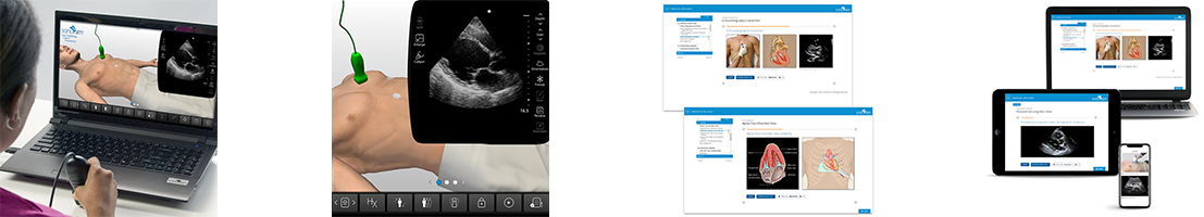
Learn the basic principles and artifacts of point of care cardiac ultrasound imaging and how to optimize image acquisition of the heart. Learn to identify and determine the size of pericardial effusion. Learn how to describe basic findings indicating right heart strain and method for determining if a pulmonary embolism is possibly present.
Course Highlights |
SonoSimulator Scanning Cases |
|
The course in this module covers topics including:
|
The 20 real patient scanning cases in this module include and cover:
|
Deep Vein Thrombosis ( DVT ) Lower Extremity Clinical Ultrasound Training
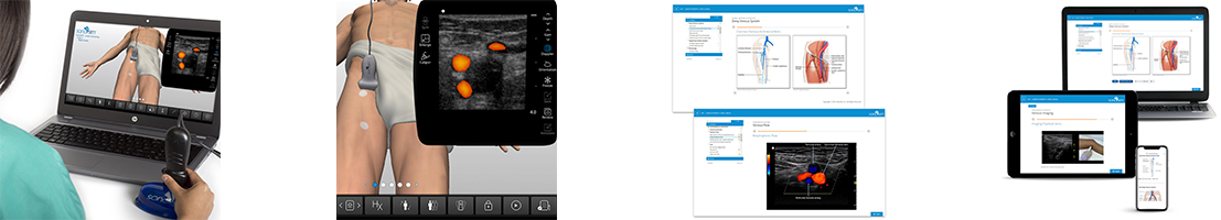
Learn how to identify and evaluate lower extremity venous anatomy with ultrasound, including imaging techniques and scanning approaches used to diagnose deep vein thrombosis (DVT). Learn how to use ultrasound to describe and evaluate a variety of patient cases involving DVT diagnoses.
Course Highlights |
SonoSimulator Scanning Cases |
|
The course in this module covers topics including:
|
The 10 real patient scanning cases in this module include and cover:
|
eFAST Protocol Clinical Training
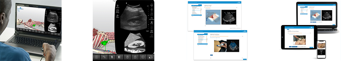
Learn the indications for administering the Focused Assessment with Sonography in Trauma (FAST) and Extended Focused Assessment with Sonography in Trauma (eFAST) examinations, as well as their goals and limitations. Learn how to obtain the five windows of the eFAST examination, including patient positioning, transducer selection, and sonographic technique. Evaluate anatomy read findings visualized in each of the five windows of the eFAST examination, which include the Right Upper Quadrant (hepatorenal), Cardiac, Left Upper Quadrant (splenorenal), Suprapubic, and Lungs.
Course Highlights |
SonoSimulator Scanning Cases |
|
The course in this module covers topics including:
|
The 10 real patient scanning cases in this module include and cover:
|
FAST Protocol Clinical Training
Learn how to obtain and describe the critical four windows of the Focused Assessment with Sonography in Trauma (FAST) examination, the current standard-of-care for emergency medicine diagnosis and management of blunt trauma. Exam overview, indications and objectives, as well as its limitations are covered, with relevant anatomy and sonographic anatomy overviews included. Practical ultrasound application factors including patient positioning, transducer selection, and sonographic technique are covered.
Course Highlights |
SonoSimulator Scanning Cases |
|
The course in this module covers topics including:
|
The 10 real patient scanning cases in this module include and cover:
|
Intestinal & Biliary Clinical Ultrasound Training
Learn how to capture, identify, and interpret ultrasound-derived imagery of the gastrointestinal system. Perform and evaluate ultrasound imaging used to screen for various gastrointestinal pathologies, including intussusception, appendicitis, cholelithiasis, and small bowel obstruction. Optimal transducer selection, imaging techniques, and scanning approaches are covered.
Course Highlights |
SonoSimulator Scanning Cases |
|
The course in this module covers topics including:
|
The 20 real patient scanning cases in this module include and cover:
|
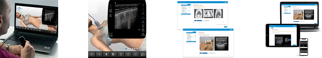
MSK Clinical Ultrasound Training
Learn how to use ultrasound to evaluate commonly encountered musculoskeletal ( MSK ) conditions. Sonographic characteristics of musculoskeletal tissues are described, as are the imaging techniques and scanning approaches utilized in MSK ultrasound.
Course Highlights |
SonoSimulator Scanning Cases |
|
The course in this module covers topics including:
|
The 10 real patient scanning cases in this module include and cover:
|
OBGYN Clinical Ultrasound Training
Learn how to use sonography to evaluate first-trimester pregnancies and basic gynecologic conditions.
Course Highlights |
SonoSimulator Scanning Cases |
|
The course in this module covers topics including:
|
The 20 real patient scanning cases in this module include and cover:
|
Ocular Clinical Ultrasound Training
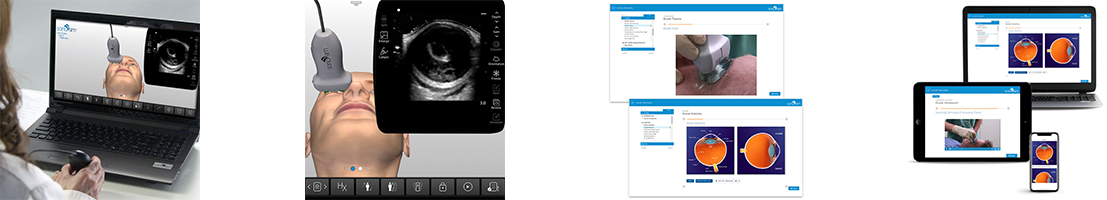
Learn how to capture and evaluate the eye with ultrasound imagery including general anatomy viewed sonographically, ultrasound techniques, and scanning approaches. Learn how to evaluate and diagnose various ocular conditions sonographically, including ocular trauma, retinal detachment, and vitreous detachment.
Course Highlights |
SonoSimulator Scanning Cases |
|
The course in this module covers topics including:
|
The 10 real patient scanning cases in this module include and cover:
|
Pulmonary Clinical Ultrasound Training
Learn how to use sonography to assess a variety of normal and pathologic pulmonary conditions, including pneumothorax, pleural effusion, alveolar interstitial syndrome, and more.
Course Highlights |
SonoSimulator Scanning Cases |
|
The course in this module covers topics including:
|
The 10 real patient scanning cases in this module include and cover:
|
Renal Clinical Ultrasound Training
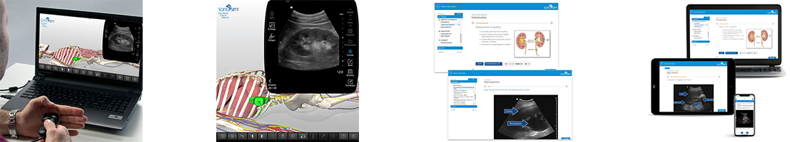
Learn how to use ultrasound to evaluate commonly encountered renal conditions, including ureteral obstruction, hydronephrosis, renal cysts, polycystic kidney, intrarenal stones, and renal tumors. Sonographic characteristics of the anatomy of the kidneys, ureters and bladder are described, as are the imaging techniques and scanning approaches utilized in renal ultrasound.
Course Highlights |
SonoSimulator Scanning Cases |
|
The course in this module covers topics including:
|
The 10 real patient scanning cases in this module include and cover:
|
RUSH Protocol Clinical Ultrasound Training
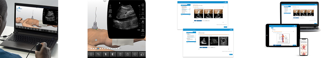
Learn how to perform the Rapid Ultrasound in Shock (RUSH) Protocol, including the anatomy and potential findings visualized in the components of the RUSH exam known as the “pump,” “tank,” and “pipes.” Learn sonographic techniques and scanning approaches used when administering the RUSH protocol.
Course Highlights |
SonoSimulator Scanning Cases |
|
The course in this module covers topics including:
|
The 10 real patient scanning cases in this module include and cover:
|
Soft Tissue Clinical Ultrasound Training
Learn how to use sonography to assess the tissues of the body, including subcutaneous tissue, fascia, muscle, tendon, vasculature, and bone. This module includes optimal transducer selection, imaging techniques, and scanning approaches for sonographically evaluating normal and pathological soft tissue. The sonographic appearance of abscesses and how they can be distinguished from cellulitis is discussed, as well as the use of ultrasound in localizing and removing foreign bodies from soft tissue.
Course Highlights |
SonoSimulator Scanning Cases |
|
The course in this module covers topics including:
|
The 10 real patient scanning cases in this module include and cover:
|


FoCUS Advanced Clinical Ultrasound Training Part 1
Learn how to obtain and describe the appropriate cardiac imaging windows for the Focused Cardiac Ultrasound (FoCUS) examination to assess the heart and surrounding vascular system. Learn where, when, and how to use Doppler adjuncts to complete a FoCUS assessment. Learn to identify and evaluate unique sonographic characteristics of the heart to assess and obtain actionable measurements for cardiac output and pulmonary artery pressure.
Course Highlights |
SonoSimulator Scanning Cases |
|
The course in this module covers topics including:
|
The 10 real patient scanning cases in this module include and cover:
|
FoCUS Advanced Clinical Ultrasound Training Part 2
Learn how to identify and evaluate the unique sonographic characteristics of valvular regurgitant and stenotic disorders of the heart as part of Focused Cardiac Ultrasound ( FoCUS ). Learn the ideal echocardiographic windows to use to detect valvular heart disorders. Learn to evaluate specific valvular disorders affecting the aortic, mitral, pulmonic, and tricuspid valves using ultrasound.
Course Highlights |
SonoSimulator Scanning Cases |
|
The course in this module covers topics including:
|
The 10 real patient scanning cases in this module include and cover:
|
LVAD Problem-Focused Echocardiography Advanced Clinical Ultrasound Training
Learn the role of bedside echocardiography in the evaluation of a Left Ventricular Assist Device ( LVAD ) patient with acute clinical decompensation. Explore the mechanics of various LVAD types, as well as LVAD complications and how to determine underlying etiology. Problem-focused echocardiographic technique is described for assessing the initial evaluation and management of LVAD patients. Develop the requisite cognitive task awareness and visuospatial skills required to perform LVAD problem-focused echocardiography.
Course Highlights |
SonoSimulator Scanning Cases |
|
The course in this module covers topics including:
|
The 3 real patient scanning cases in this module include and cover:
|
Resuscitative Transesophageal Echocardiography (TEE) Advanced Clinical Ultrasound Training
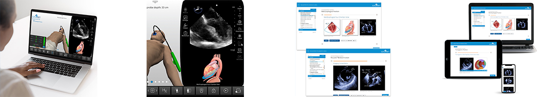
Learn about the use of transesophageal echocardiography (TEE) in the resuscitation of a patient in cardiac arrest. Resuscitative TEE indications, regional anatomy, echocardiographic anatomy, echocardiographic technique, and complications will be discussed. The course also reviews imaging tips, possible complications and risks, and benefits of resuscitative TEE. Develop the requisite cognitive task awareness and visuospatial skills foundation to build upon when learning how to perform TEE for resuscitation of patients in cardiac arrest.
Course Highlights |
SonoSimulator Scanning Cases |
|
The course in this module covers topics including:
|
The 2 real patient scanning cases & 2 SkillBox cases in this module include and cover:
|
Vascular Mapping for Preoperative Planning of Dialysis Access Advanced Clinical Ultrasound Training
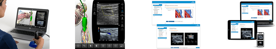
Learn how to use ultrasound to perform vascular mapping as part of preoperative planning for dialysis access. Explore the use of ultrasound to survey upper extremity vasculature and determine the most desirable hemodialysis access site for arteriovenous fistula or graft creation. Relevant anatomy of the upper extremity vasculature, imaging protocols, and the various types of arteriovenous fistulae and grafts are discussed. Develop the requisite cognitive task awareness and psychomotor skills required for performing ultrasound vascular mapping of the upper extremities.
Course Highlights |
SonoSimulator Scanning Cases |
|
The course in this module covers topics including:
|
The 10 real patient scanning cases in this module include and cover:
|
Neonatal & Infant Neurosonography Advanced Clinical Ultrasound Training
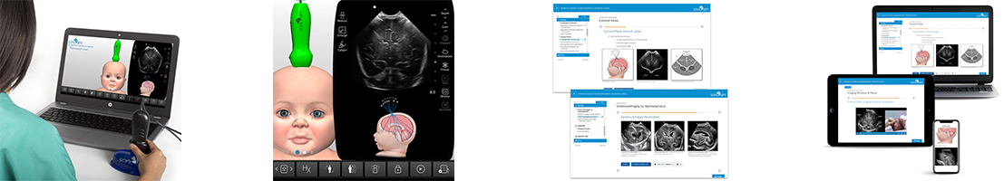
Learn how to perform neonatal and infant neurosonography. Get a thorough overview of the neuroanatomy of preterm and term neonates and infants, and explores the clinical indications for sonographic examination, an in-depth review of sonographic neuroanatomy and imaging techniques, and an introduction to several more commonly encountered pathologic conditions. Develop the requisite cognitive task awareness and visuospatial skills required to perform neonatal and infant neurosonography.
Course Highlights |
SonoSimulator Scanning Cases |
|
The course in this module covers topics including:
|
The 10 real patient scanning cases in this module include and cover:
|
Pediatric Lung Sonography Advanced Clinical Ultrasound Training
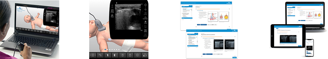
Learn how to use sonography to evaluate pediatric patients presenting with a variety of normal and pathologic pulmonary conditions. The sonographic characteristics of the more commonly encountered pediatric respiratory conditions will be discussed, with special emphasis on diseases of neonates and infants. Review relevant anatomy and explore the role of sonography in the evaluation of respiratory conditions such as: pneumonia, pleural effusion, pneumothorax, atelectasis, transient tachypnea, meconium aspiration syndrome, respiratory distress syndrome, bronchiolitis, and bronchopulmonary dysplasia. Develop the requisite cognitive task awareness and visuospatial skills required to perform sonographic examination of pediatric patients presenting with respiratory conditions.
Course Highlights |
SonoSimulator Scanning Cases |
|
The course in this module covers topics including:
|
The 10 real patient scanning cases in this module include and cover:
|
First-Trimester Pregnancy Advanced Clinical Ultrasound Training
Review traditional illustrative anatomy of the female pelvis and embryologic development alongside sonographic anatomy. Learn appropriate imaging techniques used to identify and characterize anatomic components of the female anatomy during the first-trimester of pregnancy. Learn to identify and interpret anatomy in ultrasound imaging. Identify and evaluate the unique sonographic characteristics of this phase of pregnancy, including pathology and pregnancy failure.
Course Highlights |
SonoSimulator Scanning Cases |
|
The course in this module covers topics including:
|
The 10 real patient scanning cases in this module include and cover:
|
Second and Third Trimester Pregnancy Advanced Clinical Ultrasound Training Part 1
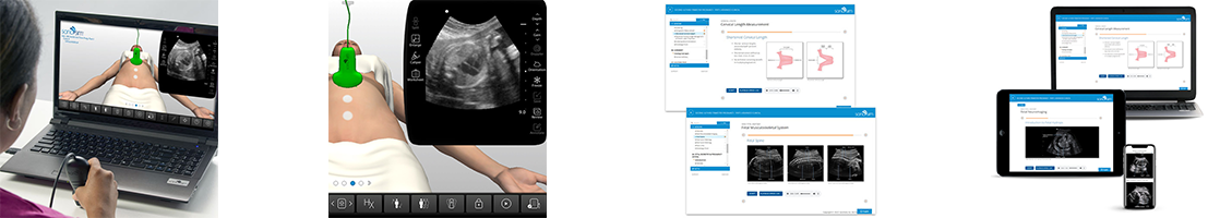
Get an in-depth review of the sonographic evaluation and monitoring of normal embryologic and fetal development, including multiple pregnancies, and pathologic conditions that become evident in the second- and third-trimester of pregnancy. Develop the requisite hands-on psychomotor skills and cognitive task awareness (e.g. fetal biometry) required to optimally perform second and third trimester pregnancy ultrasound.
Course Highlights |
SonoSimulator Scanning Cases |
|
The course in this module covers topics including:
|
The 10 real patient scanning cases in this module include and cover:
|
Second and Third Trimester Pregnancy Advanced Clinical Ultrasound Training Part 2
Get an in-depth review reinforcing the use of fetal biometry to evaluate fetal development of singleton and multiple pregnancies, weight discrepancies, amniotic fluid volume indexes, and maximal vertical pocket identification during the second- and third-trimesters of pregnancy. Sonographic assessment of fetal presentation, placenta, and placental location are discussed. Pathologic conditions such as intrauterine growth restriction and fetal macrosomia are described in detail. Develop the requisite hands-on psychomotor skills and cognitive task awareness required to optimally perform second and third trimester pregnancy ultrasound.
Course Highlights |
SonoSimulator Scanning Cases |
|
The course in this module covers topics including:
|
The 10 real patient scanning cases in this module include and cover:
|
GYN Ultrasound of Nonpregnant Normal Uterus Advanced Clinical Training
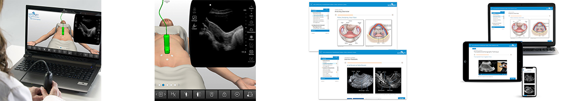
Learn to identify and evaluate a nonpregnant female uterus and pelvis using ultrasonography, including anatomy, sonographic characteristics and ultrasound imaging techniques. Learn to recognize and evaluate normal variations of uterine appearance and positioning in the nonpregnant female, with sonography.
Course Highlights |
SonoSimulator Scanning Cases |
|
The course in this module covers topics including:
|
The 10 real patient scanning cases in this module include and cover:
|
GYN Ultrasound of Nonpregnant Abnormal Uterus Advanced Clinical Training Part 1
Learn to identify and evaluate the nonpregnant uterus, including anatomy, sonographic characteristics and imaging techniques. Understand how to evaluate the abnormal, nonpregnant uterus with ultrasonography. Learn to recognize and evaluate various abnormal conditions of the myometrium in the nonpregnant female, using ultrasound.
Course Highlights |
SonoSimulator Scanning Cases |
|
The course in this module covers topics including:
|
The 10 real patient scanning cases in this module include and cover:
|
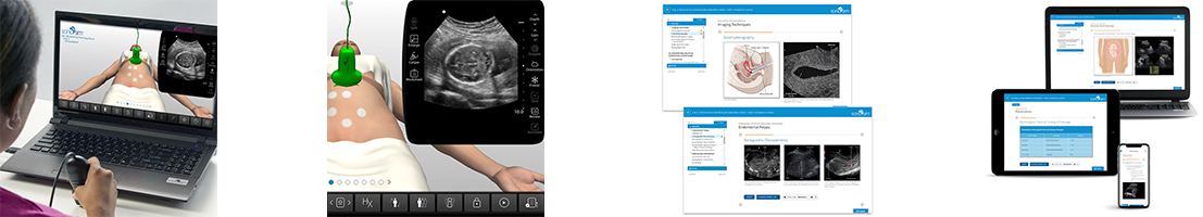
GYN Ultrasound of Nonpregnant Abnormal Uterus Advanced Clinical Training Part 2
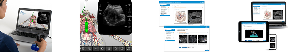
Learn to identify and evaluate various endometrial conditions in the nonpregnant abnormal uterus, including sonographic characteristics, and imaging techniques.
Course Highlights |
SonoSimulator Scanning Cases |
|
The course in this module covers topics including:
|
The 5 real patient scanning cases in this module include and cover:
|
GYN Ultrasound Normal Adnexa Advanced Clinical Training
Learn to identify and evaluate the normal adnexa, including anatomy, unique sonographic characteristics and ultrasound imaging techniques used in evaluating the normal adnexa. Learn to identify and evaluate normal variations of appearance and positioning of the normal adnexa.
Course Highlights |
SonoSimulator Scanning Cases |
|
The course in this module covers topics including:
|
The 10 real patient scanning cases in this module include and cover:
|
GYN Ultrasound Nonmalignant Adnexal Conditions Advanced Clinical Training
Learn to evaluate adnexal masses using ultrasound, including imaging techniques used in sonographic evaluation of nonmalignant adnexal conditions. Understand the implications of ultrasound findings for specific gynecological conditions. Learn to identify and evaluate nonmalignant adnexal conditions, such as hydrosalpinx, tubo-ovarian abscess, and adnexal torsion, using ultrasonography.
Course Highlights |
SonoSimulator Scanning Cases |
|
The course in this module covers topics including:
|
The 10 real patient scanning cases in this module include and cover:
|
GYN Ultrasound Malignant Adnexal Conditions Advanced Clinical Training
Learn to evaluate adnexal masses using ultrasound, including imaging techniques used in sonographic evaluation of malignant adnexal conditions. Learn to identify the risk factors for ovarian cancer and evaluate ovarian cancer via ultrasonography.
Course Highlights |
SonoSimulator Scanning Cases |
|
The course in this module covers topics including:
|
The 10 real patient scanning cases in this module include and cover:
|


Ultrasound-Guided Procedures Introductory Training
Learn the basic principles of performing ultrasound-guided procedures. Develop the requisite hands-on psychomotor skills and cognitive task awareness required to optimally perform ultrasound-guided procedures. Included SkillBox scanning cases provide risk-free cognitive task training opportunities.
Course Highlights |
SonoSimulator Scanning Cases |
|
The course in this module covers topics including:
|
The 10 SkillBox scanning cases in this module include:
|
Peripheral Venous Access Ultrasound-Guided Procedure Training
Learn how to use sonography to guide peripheral venous access in patients. The needle-based peripheral venous access procedure feature provides cognitive task training on ultrasound-guided needle-based procedures. Ultrasound-guided peripheral venous access procedure indications, sonographic techniques and scanning approaches, including imaging tips, pitfalls, and procedure complications are described.
Course Highlights |
SonoSimulator Scanning Cases |
|
The course in this module covers topics including:
|
The 10 real patient scanning cases in this module include and cover:
|
Femoral Line Placement Ultrasound-Guided Procedure Training
Get an in-depth understanding of the basic principles of how to perform ultrasound-guided femoral central line placement. Develop the requisite hands-on psychomotor skills and cognitive task awareness required to optimally perform femoral vein access with the aid of ultrasound.
Course Highlights |
SonoSimulator Scanning Cases |
|
The course in this module covers topics including:
|
The 10 real patient scanning cases in this module include and cover:
|
Internal Jugular Vein Cannulation Ultrasound-Guided Procedure Training
Get an in-depth understanding of the basic principles of how to perform ultrasound-guided internal jugular vein cannulation. Develop the requisite hands-on psychomotor skills and cognitive task awareness required to optimally perform ultrasound-guided internal jugular vein cannulation. Ultrasound-guided internal jugular vein cannulation procedure indications, sonographic techniques and scanning approaches, including imaging tips, pitfalls, and procedure complications are also provided.
Course Highlights |
SonoSimulator Scanning Cases |
|
The course in this module covers topics including:
|
The 10 real patient scanning cases in this module include and cover:
|
Subclavian Vein Cannulation Ultrasound-Guided Procedure Training
Get in-depth understanding of the basic principles of how to perform ultrasound-guided subclavian vein cannulation. Develop the requisite hands-on psychomotor skills and cognitive task awareness required to optimally perform ultrasound-guided subclavian vein cannulation. Ultrasound-guided subclavian vein cannulation procedure indications, sonographic techniques and scanning approaches, including imaging tips, pitfalls, and procedure complications are also provided.
Course Highlights |
SonoSimulator Scanning Cases |
|
The course in this module covers topics including:
|
The 10 real patient scanning cases in this module include and cover:
|
Ultrasound-Guided Paracentesis Procedure Training
Learn how to perform an ultrasound-guided paracentesis. This course explores the use of ultrasound to detect ascites, regional anatomy, clinical indications for performing paracentesis, procedural complications, and procedural steps for performing ultrasound-guided paracentesis. Develop the requisite cognitive task awareness and visuospatial skills required to perform ultrasound-guided paracentesis.
Course Highlights |
SonoSimulator Scanning Cases |
|
The course in this module covers topics including:
|
The 10 real patient cases in this module include and cover:
|
Ultrasound-Guided Pericardiocentesis Procedure Training
Learn the principles of performing an ultrasound-guided pericardiocentesis. Explore the use of ultrasound to detect pericardial effusions, understand clinical indications for performing pericardiocentesis, and how to identify echocardiographic findings suggestive of cardiac tamponade physiology. Learn the steps necessary for performing ultrasound-guided pericardiocentesis. Develop the requisite cognitive task awareness and visuospatial skills required to perform ultrasound-guided pericardiocentesis.
Course Highlights |
SonoSimulator Scanning Cases |
|
The course in this module covers topics including:
|
The 10 real patient scanning cases in this module include and cover:
|
Ultrasound-Guided Thoracentesis Procedure Training
Learn how to perform an ultrasound-guided thoracentesis. The course explores the use of ultrasound to detect pleural effusions, clinical indications for performing a thoracentesis, procedural complications, and procedural steps for performing ultrasound-guided thoracentesis. Develop the requisite cognitive task awareness and visuospatial skills required to perform ultrasound-guided thoracentesis.
Course Highlights |
SonoSimulator Scanning Cases |
|
The course in this module covers topics including:
|
The 10 real patient scanning cases in this module include and cover:
|
Biceps Tendon Sheath Injection Ultrasound-Guided Procedure Training
Learn both the regional and sonographic anatomy and how to interpret ultrasound imagery of the shoulder joint. Learn sonographic techniques and scanning approaches used in ultrasound-guided biceps tendon sheath injection. Learn to evaluate various biceps tendon sheath pathologies with ultrasonography and clinical indications for performing biceps tendon sheath injections.
Course Highlights |
SonoSimulator Scanning Cases |
|
The course in this module covers topics including:
|
The real adult patient training case in this module includes and covers:
|
Plantar Fasciitis Ultrasound-Guided Injection Procedures
Learn the basic principles of performing ultrasound-guided plantar intrafascial & perifascial injections. Review relevant anatomic landmarks and the use of ultrasound to evaluate the plantar fascia. Learn commonly agreed upon indications for performing this procedure. Develop cognitive task awareness and visuospatial skills required to perform ultrasound-guided injections to alleviate plantar fasciitis.
Course Highlights |
SonoSimulator Scanning Cases |
|
The course in this module covers topics including:
|
The 2 real patient scanning cases in this module include and cover:
|
Acromioclavicular Joint Injection & Aspiration Ultrasound-Guided Procedure Training
Learn both the regional and sonographic anatomy of the acromioclavicular joint and how to interpret ultrasound imagery of this joint. Learn sonographic techniques and scanning approaches used in ultrasound-guided acromioclavicular joint injection and aspiration. Learn to evaluate various acromioclavicular joint pathologies with ultrasonography and clinical indications for performing acromioclavicular joint injections and acromioclavicular joint aspiration.
Course Highlights |
SonoSimulator Scanning Cases |
|
The course in this module covers topics including:
|
The one (1) real patient scanning case in this module includes and covers:
|
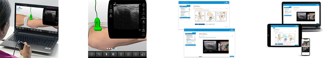
Glenohumeral Joint Injection & Aspiration Ultrasound-Guided Procedure Training
Learn the relationship between traditional illustrative anatomy and sonographic anatomy of the glenohumeral joint. Learn how glenohumeral joint injections and glenohumeral joint aspiration is performed, including the location and appearance of critical anatomical components in ultrasound imaging. Sonographic techniques and scanning approaches used in performing ultrasound-guided glenohumeral joint injection and aspiration procedures are covered. Ultrasound-guided glenohumeral joint injection & aspiration procedure indications, sonographic techniques and scanning approaches, including imaging tips, pitfalls, and procedure complications are covered.
Course Highlights |
SonoSimulator Scanning Cases |
|
The course in this module covers topics including:
|
The real patient training case in this module includes and covers:
|
Subacromial-Subdeltoid Bursa Injection & Aspiration Ultrasound-Guided Procedure Training
Get an in-depth understanding of the basic principles of how to perform ultrasound-guided subacromial-subdeltoid bursal injection and aspiration. The course explores the use of ultrasound to detect bursal fluid and clinical indications for performing the procedure. Subacromial-subdeltoid bursitis, subacromial impingement syndrome, and rotator cuff injuries are discussed. Develop the requisite cognitive task awareness and visuospatial skills required to perform ultrasound-guided subacromial-subdeltoid bursa injection and aspiration. Ultrasound-guided subacromial-subdeltoid bursa injection and aspiration procedure indications, sonographic techniques and scanning approaches, including imaging tips, pitfalls, and procedure complications are described.
Course Highlights |
SonoSimulator Scanning Cases |
|
The course in this module covers topics including:
|
The real patient scanning case in this module includes and covers:
|
Tibiotalar Joint Injection & Aspiration Ultrasound-Guided Procedure Training
Get an in-depth understanding of the basic principles of how to perform ultrasound-guided tibiotalar joint injection and/or aspiration. Review relevant anatomy and explore the use of ultrasound to evaluate tibiotalar joint pathologies and clinical indications for performing the procedure. Acute and chronic pathologic conditions that afflict the tibiotalar joint are discussed, including trauma, degenerative arthritis, cartilaginous lesions, inflammatory conditions, and infections. Develop the requisite cognitive task awareness and visuospatial skills required to perform ultrasound-guided tibiotalar joint injection.
Course Highlights |
SonoSimulator Scanning Cases |
|
The course in this module covers topics including:
|
The real patient training case in this module includes and covers:
|
Metatarsophalangeal Joint Injection & Aspiration Ultrasound-Guided Procedure Training
Learn the basic principles of performing ultrasound-guided metatarsophalangeal joint injection and aspiration. Review relevant anatomy and the use of ultrasound to evaluate metatarsophalangeal joint pathologies. Learn the clinical indications for performing the procedure. Review acute and chronic pathologic conditions that afflict the metatarsophalangeal joint, including trauma, degenerative arthritis, inflammatory conditions, infections, and masses. Develop cognitive task awareness and visuospatial skills required to perform ultrasound-guided metatarsophalangeal joint injection.
Course Highlights |
SonoSimulator Scanning Cases |
|
The course in this module covers topics including:
|
The 2 real patient scanning cases in this module include and cover:
|
Neonatal & Infant Lumbar Puncture Ultrasound-Guided Procedure Training
Gain an in-depth understanding of the basic principles of performing ultrasound-guided neonatal and infant lumbar punctures. Review relevant anatomic landmarks that are instrumental to performing ultrasound-guided lumbar punctures. Develop cognitive task awareness and visuospatial skills required to perform the procedure to evaluate for sepsis or central nervous system infections. Review the anatomy of the neonatal and infant spine, and commonly agreed-upon clinical indications for performing ultrasound-guided lumbar punctures. Learn optimal transducer selection, patient positioning, imaging approaches, and sonographic techniques for procedure execution. Imaging tips and pitfalls, and potential procedure complications are discussed.
Course Highlights |
SonoSimulator Scanning Cases |
|
The course in this module covers topics including:
|
The 2 real patient scanning cases in this module include and cover:
|


