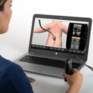
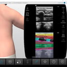
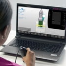
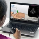
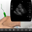
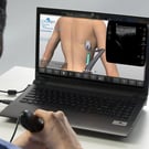
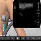
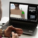
Using real-time ultrasound guidance for a multitude of procedures, such as peripheral IV access, is becoming the “standard of care”. However, performing these challenging procedures requires significant ultrasound procedure knowledge and psychomotor skills.
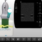
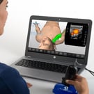
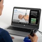
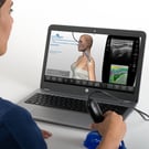
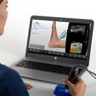
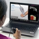
SonoSim provides a safe and convenient setting for learners to practice a variety of ultrasound-guided procedures. This unrestricted access to deliberate practice is an essential element for acquiring the ultrasound procedure knowledge, psychomotor skill, and confidence required for success with ultrasound procedures.


.jpg?width=135&height=135&name=procedure_methatarsophalangeal_case_screen__08737%20(1).jpg)