Vascular Ultrasound Training

Hands-On Vascular Ultrasound Training for an Immersive Learning Experience

Vascular Ultrasound & Advanced Doppler Imaging

Courses & Guided Scanning Practice

SonoSim's hands-on vascular ultrasound training material is expertly designed to bolster the skills and knowledge of learners in vascular ultrasound, including Doppler ultrasound applications. Our curriculum is authored by leading experts in vascular access ultrasound, peer-reviewed, and CME-accredited. Every ultrasound training topic includes a course, virtual scanning cases with Doppler simulation, knowledge assessments, scanning assignments, guided imaging protocols, and a Mastery Exam. Participants have the opportunity to earn certificates, build their vascular ultrasound image portfolios, and claim CME credits when needed. Our in-depth learning modules serve as an invaluable reference guide for those interested in ultrasound vascular studies.

SonoSim vascular ultrasound training courses, SonoSimulator® hands-on virtual scanning cases, and imaging protocols are designed to adhere to broadly accepted consensus guidelines (e.g., AIUM). The DMS Imaging Protocols are specifically designed to match professional society guidelines. SonoSim vascular ultrasound training content is designed to assist DMS programs prepare their students to pass the ARDMS Vascular Sonography exam.

Understanding how to perform vascular ultrasound is critically important across multiple medical specialties. Doppler ultrasound is central to performing vascular ultrasound examinations. SonoSim provides a series of training courses and virtual hands-on scanning cases that starts with learning how to use vascular ultrasound to scan normal anatomy. 

The Fundamentals of Ultrasound Course coupled with the SPI study tool helps develop a solid foundation of ultrasound physics that supports future vascular ultrasound learning. Advanced courses and cases further develop vascular ultrasound skills, specifically by applying Doppler ultrasound imaging towards a diverse series of applications. DMS imaging protocols provide in-depth guided training that instructs learners how to perform vascular ultrasound using Doppler, as indicated. A series of ultrasound-guided vascular access procedures are also available.
Color doppler ultrasound footage in SonoSim vascular ultrasound training
GET IN TOUCH

Each topic is meticulously structured to meet the critical learning objectives necessary for mastering vascular ultrasound. This systematic approach covers key areas such as:

  • Vascular Ultrasound Indications
  • Vascular Anatomy & Physiology
  • Vascular Sonographic Anatomy
  • Sonographic Technique for Vascular Ultrasound
  • Tips & Pitfalls When Imaging the Vascular System
  • Pathologic Vascular Case Studies

POCUS & DMS Protocol Scanning Assignments

Customized scanning assignments are provided based upon whether learners are pursuing diagnostic medical sonography (DMS) or point-of-care ultrasound (POCUS) training.

DMS Protocol Scanning Assignments are available for many topics/protocols.


Hands-on Vascular Ultrasound Doppler Training

Enhance and Broaden Your Capabilities with Our Comprehensive Vascular Access Ultrasound Course

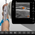
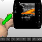
Our all-encompassing training platform fuels every learner's journey toward mastering hands-on vascular ultrasound training in diverse clinical situations. Learners get the chance to practice acquiring & interpreting vascular Doppler ultrasound images & identifying vascular anomalies.

SonoSim cloud-based, multimedia, didactic courses are created by leading ultrasound experts and are internet accessible immediately after purchase on any device. There are knowledge checks with real-time feedback throughout each course, as well as a Mastery Test that is automatically graded.

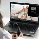
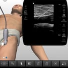
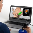
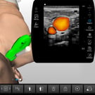
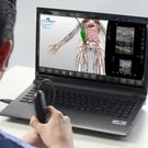
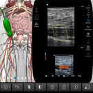
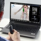
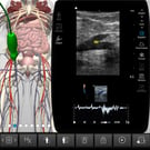
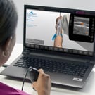
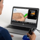
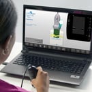
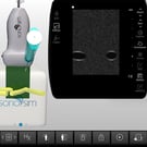
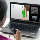
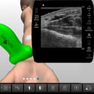
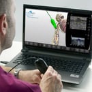
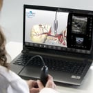
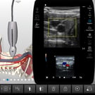
The SonoSimulator® helps develop and maintain the critical visuomotor and visuospatial skills that are central to image acquisition and interpretation with real patient imagery, expert tutorials on-demand, and real-time feedback on success.

Aorta & Inferior Vena Cava ( IVC ) Anatomy & Physiology Ultrasound Training

Image Carrousel 5

Learn the relationship of traditional illustrative and sonographic anatomy of the aorta and inferior vena cava (IVC) and how to interpret ultrasound imaging of the aorta & IVC. Learn sonographic techniques and scanning approaches used in ultrasound imaging to assess the aorta and inferior vena cava. Learn to identify and characterize the aorta / IVC with ultrasonography.

Course Highlights

SonoSimulator Scanning Cases

The course in this module covers topics including:

  • Anatomy and Physiology of the Aorta and Inferior Vena Cava
  • Sonographic Anatomy of the Aorta & Inferior Vena Cava
  • Sonographic Techniques including Doppler Adjuncts
  • Pearls & Pitfalls when Evaluating the Aorta and Inferior Vena Cava

The 5 real patient scanning cases in this module include and cover:

  • Patient Types: Adult Only, Male & Female
  • Relevant Findings/Pathology: Normal Examples of Aorta and IVC Anatomy
  • Doppler & Imaging Modes: Color-Flow Doppler, Pulsed-Wave Doppler
  • Transducer Types: Curvilinear
  • Special Case Features: Additional Abdominal CT imaging, Anatomic Layer Removal

How to buy5


Aorta & Inferior Vena Cava ( IVC ) Clinical Ultrasound Training

Image Carrousel 6

Learn how to evaluate aortic anatomy and identify abdominal aortic aneurysms (AAAs) with ultrasound. How to evaluate IVC anatomy and how to estimate central venous pressure (CVP) are also discussed. Practical application factors including transducer selection, imaging techniques, and scanning approaches are covered.

Course Highlights

SonoSimulator Scanning Cases

The course in this module covers topics including:

  • Ultrasound Applications for Abdominal Aortic Aneurysms (AAAs)
  • Aortic Anatomy
  • Sonography Anatomy & Characteristics
  • Transducer Selection & Settings
  • Patient Positioning
  • 6 AAA Case Studies, as well as Cases of Aortic Dissection, Saccular Aneurysm and AAA with a Graft
  • Inferior Vena Cava ( IVC ) Evaluation
  • Central Venous Pressure ( CVP ) Estimation
  • Tips, Pitfalls, & Complications

The 20 real patient scanning cases in this module include and cover:

  • Patient Types: Male & Female; 17 Adult & 3 Pediatric
  • Relevant Findings/Pathology: Normal Adult & Pediatric Patients, Abdominal Aortic Aneurysm Patients, Aortic Aneurysm with Mural Thrombus Patient, Aortic Aneurysm with Endovascular Stent Patient, Fusiform Abdominal Aneurysm Patient, Hypovolemic IVC Caval Index Patient, Normovolemic IVC Caval Index Patient, Hypervolemic IVC Caval Index Patient
  • Doppler & Imaging Modes: Color-Flow Doppler, Power Doppler, Pulse-Wave Doppler
  • Transducer Types: Linear, Curvilinear, Phased Array
  • Special Case Features: Abdominal CT Imaging, Anatomic Layer Removal

How to buy5


Arm Arterial Anatomy & Physiology Ultrasound Training

Image Carrousel 7

Learn the relationship of traditional illustrative and sonographic anatomy of the arm and how to interpret ultrasound imaging of the upper extremity arterial system. Learn sonographic techniques and scanning approaches used in ultrasound imaging of the upper extremity arterial system. Learn to identify and characterize the macroscopic and regional anatomical components of the upper extremity arterial system with ultrasonography.

Course Highlights

SonoSimulator Scanning Cases

The course in this module covers topics including:

  • Upper Extremity Arterial Anatomy
  • Arm Arterial Physiology Including Circulation & Blood Flow
  • Sonographic Anatomy of Upper Extremity Arteries
  • Sonographic Techniques
  • Pearls & Pitfalls when Evaluating the Arteries

The 5 real patient scanning cases in this module include and cover:

  • Patient Types: Adult Only, Male & Female
  • Relevant Findings/Pathology: Normal Examples of Arterial Arm Anatomy
  • Doppler & Imaging Modes: Color-Flow Doppler, Pulsed-Wave Doppler
  • Transducer Types: Linear
  • Sonographic Technique
  • Special Case Features: Compression, Anatomic Layer Removal

How to buy5


Arm Venous Anatomy & Physiology Ultrasound Training

Image Carrousel 8

Learn the relationship of traditional illustrative and sonographic anatomy of the arm and how to interpret ultrasound imaging of arm veins. Learn sonographic techniques and scanning approaches used in ultrasound imaging to assess the venous system of the upper extremities. Learn to identify and characterize arm veins with ultrasonography.

Course Highlights

SonoSimulator Scanning Cases

The course in this module covers topics including:

  • Arm Vein Anatomy including Deep and Superficial Veins
  • Venous Arm Physiology including Capacity and Flow
  • Sonographic Anatomy of the Arm Veins
  • Sonographic Anatomy
  • Sonographic Techniques
  • Pearls & Pitfalls when Evaluating the Arm Veins

The 5 real patient scanning cases in this module include and cover:

  • Patient Types: Adult Only, Male & Female
  • Relevant Findings/Pathology: Normal Examples of Venous Arm Anatomy
  • Doppler & Imaging Modes: Color-Flow Doppler, Pulsed-Wave Doppler
  • Transducer Types: Linear
  • Special Case Features: Compression, Anatomic Layer Removal

How to buy5


Cerebrovascular Anatomy & Physiology Ultrasound Training

Image Carrousel 16

Learn the relationship of traditional illustrative and sonographic anatomy of the cerebral vasculature and how to interpret cerebrovascular ultrasound imaging. Learn sonographic techniques and scanning approaches used in ultrasound imaging to assess cerebral vasculature. Learn to identify and characterize cerebral vasculature with ultrasonography.

Course Highlights

SonoSimulator Scanning Cases

The course in this module covers topics including:

  • Cerebral Blood Supply
  • Cerebrovascular Physiology
  • Histophysiology
  • Cerebrovascular Sonographic Anatomy
  • Sonographic Techniques for scanning Carotid & Vertebral Arteries & the Internal Jugular Vein
  • Pearls & Pitfalls when Evaluating the Cerebrovascular System

The 5 real patient scanning cases in this module include and cover:

  • Patient Types: Adult Only, Male & Female
  • Relevant Findings/Pathology: Normal Examples of Cerebrovascular Anatomy
  • Doppler & Imaging Modes: Color-Flow Doppler, Pulsed-Wave Doppler
  • Transducer Types: Linear
  • Special Case Features: Additional Cerebrovascular MRA Imaging, Anatomic Layer Removal

How to buy5


Deep Vein Thrombosis ( DVT ) Lower Extremity Clinical Ultrasound Training

Image Carrousel 17

Learn how to identify and evaluate lower extremity venous anatomy with ultrasound, including imaging techniques and scanning approaches used to diagnose deep vein thrombosis (DVT). Learn how to use ultrasound to describe and evaluate a variety of patient cases involving DVT diagnoses.

Course Highlights

SonoSimulator Scanning Cases

The course in this module covers topics including:

  • Normal Venous System Anatomy and Physiology
  • Sonographic Venous Anatomy & Flow
  • Sonographic Technique & Examination
  • POCUS DVT Examination
  • Possible Diagnoses including DVT & Alternative Conditions

The 10 real patient scanning cases in this module include and cover:

  • Patient Types: Adult Only, Male & Female
  • Relevant Findings/Pathology: Partially occlusive thrombi cases; Completely Occlusive Thrombi Cases; Acute Thrombi Cases; Chronic-Appearing Thrombi Cases; Identified Thrombi in the Common Femoral, Proximal Femoral, Profunda Femoris, Mid-Femoral, and Popliteal Venous Segments of the Leg; Scan DVT Differential Diagnosis Conditions (e.g., Baker’s Cyst)
  • Doppler & Imaging Modes: Color-Flow Doppler, Pulsed-Wave Doppler, Power Doppler
  • Scanning Approaches: Groin, Upper thigh, Mid thigh, Popliteal fossa
  • Transducer Types: Linear
  • Special Case Features: Compression

How to buy5


Femoral Line Placement Ultrasound-Guided Procedure Training

Image Carrousel 22

Get an in-depth understanding of the basic principles of how to perform ultrasound-guided femoral central line placement. Develop the requisite hands-on psychomotor skills and cognitive task awareness required to optimally perform femoral vein access with the aid of ultrasound.

Course Highlights

SonoSimulator Scanning Cases

The course in this module covers topics including:

  • Procedure Indications and Contraindications
  • Site Selection
  • Sonographic Anatomy & Characteristics
  • Transducer Selection & Settings
  • Ergonomic Considerations
  • Patient Positioning
  • Infection Prevention
  • Survey Scan and Procedure Steps
  • Doppler-Mode Imaging
  • Tips, Pitfalls, and Complications

The 10 real patient scanning cases in this module include and cover:

  • Patient Types: Adult Only, Male & Female
  • Relevant Findings/Pathology: Normal Patient Anatomy, Variable Body Habitus, Variable Hemodynamic and Hydration Status
  • Doppler & Imaging Modes: Power Doppler
  • Special Case Features: Compressions

How to buy5


Internal Jugular Vein Cannulation Ultrasound-Guided Procedure Training

Image Carrousel 38

Get an in-depth understanding of the basic principles of how to perform ultrasound-guided internal jugular vein cannulation. Develop the requisite hands-on psychomotor skills and cognitive task awareness required to optimally perform ultrasound-guided internal jugular vein cannulation. Ultrasound-guided internal jugular vein cannulation procedure indications, sonographic techniques and scanning approaches, including imaging tips, pitfalls, and procedure complications are also provided.

Course Highlights

SonoSimulator Scanning Cases

The course in this module covers topics including:

  • Procedure Indications & Contraindications
  • Site Selection
  • Sonography Anatomy & Characteristics
  • Transducer Selection & Settings
  • Ergonomic Considerations
  • Patient Positioning
  • Infection Prevention
  • Survey Scan & Procedure Steps
  • Doppler-Mode Imaging
  • Tips, Pitfalls, & Complications

The 10 real patient scanning cases in this module include and cover:

  • Patient Types: Adult Only, Male & Female
  • Relevant Findings/Pathology: Variable Body Morphologies, Right and Left Neck, Variable Hemodynamic & Hydration Status
  • Doppler & Imaging Modes: Power Doppler
  • Scanning Approaches: Transverse & Longitudinal
  • Procedural Techniques: In-Plane & Out-of-Plane Needle Guidance
  • Transducer Types: Linear
  • Special Case Features: Compression, Anatomic Layer Removal

How to buy5


Leg Arterial Anatomy & Physiology Ultrasound Training

Image Carrousel 41

Learn how to use ultrasound in the evaluation of the lower extremity arterial system, including lower extremity arterial anatomy and physiology and sonographic anatomy of the lower extremity arterial system. Optimal transducer selection, patient positioning, imaging approaches, and techniques for sonographically evaluating the lower extremity arterial system are discussed.

Course Highlights

SonoSimulator Scanning Cases

The course in this module covers topics including:

  • Arterial Anatomy & Physiology of the Leg
  • Sonographic Anatomy of Leg Arteries including Doppler Adjuncts
  • Sonographic Imaging Techniques of the Lower Extremities
  • Pearls & Pitfalls when Evaluating the Leg Arteries

The course in this module covers topics including:

  • Arterial Anatomy & Physiology of the Leg
  • Sonographic Anatomy of Leg Arteries including Doppler Adjuncts
  • Sonographic Imaging Techniques of the Lower Extremities
  • Pearls & Pitfalls when Evaluating the Leg Arteries

How to buy5


Leg Venous Anatomy & Physiology Ultrasound Training

Image Carrousel 42

Learn how to use ultrasound in the evaluation of the lower extremity vasculature, including lower extremity venous anatomy and physiology, and sonographic anatomy of the lower extremity venous system. Optimal transducer selection, patient positioning, imaging approaches, and techniques for sonographically evaluating the lower extremity venous system are discussed.

Course Highlights

SonoSimulator Scanning Cases

The course in this module covers topics including:

  • Anatomy of the Venous System of the Leg including Deep and Superficial Veins
  • Venous Leg Physiology including Capacity and Flow
  • Sonographic Anatomy of Leg Veins
  • Sonographic Imaging Techniques for Hip, Thigh, Calf, Ankle & Foot Veins
  • Pearls & Pitfalls when Evaluating Leg Veins

The 5 real patient scanning cases in this module include and cover:

  • Patient Types: Male & Female, 4 Adult & 1 Pediatric
  • Relevant Findings/Pathology: Normal Examples of Venous Leg Anatomy
  • Doppler & Imaging Modes: Color-Flow Doppler, Pulsed-Wave Doppler
  • Transducer Types: Linear, Curvilinear
  • Special Case Features: Compression, Anatomic Layer Removal

How to buy5


Peripheral Venous Access Ultrasound-Guided Procedure Training

Image Carrousel 54

Learn how to use sonography to guide peripheral venous access in patients. The needle-based peripheral venous access procedure feature provides cognitive task training on ultrasound-guided needle-based procedures. Ultrasound-guided peripheral venous access procedure indications, sonographic techniques and scanning approaches, including imaging tips, pitfalls, and procedure complications are described.

Course Highlights

SonoSimulator Scanning Cases

The course in this module covers topics including:

  • Procedure Indications & Contraindications
  • Site Selection
  • Sonography Anatomy & Characteristics
  • Transducer Selection & Settings
  • Ergonomic Considerations
  • Patient Positioning
  • Ergonomic Considerations
  • Infection Prevention
  • Survey Scan & Procedure Steps
  • Doppler-Mode Imaging
  • Tips, Pitfalls & Complications

The 10 real patient scanning cases in this module include and cover:

  • Patient Types: Male & Female, 7 Adult & 3 Pediatric
  • Relevant Findings/Pathology: Variable Body Morphology, Right and Left Arms, Dorsal Metacarpal Veins, Cephalic Veins, Brachial Veins, Basilic Veins, Median Cubital Vein
  • Scanning Approaches: Short Axis & Long Axis
  • Doppler & Imaging Modes: Power Doppler
  • Procedure Techniques: In-Plane & Out-of-Plane Needle Guidance
  • Transducer Types: Linear
  • Special Case Features: Compression

How to buy5


Renal Transplant Ultrasound Advanced Clinical Training


Learn how to perform renal transplant ultrasound to evaluate renal allografts. The role of ultrasound & indications for renal allograft ultrasound examination is presented. Renal transplant allograft and regional anatomy are explored. Optimal sonographic technique, imaging protocols, and the sonographic characteristics of normal renal transplant allografts are presented.  

A series of case studies are used to review how ultrasound is used to identify more commonly encountered renal transplant complications. The procedural knowledge and requisite cognitive task awareness and visuospatial skills required to perform renal transplant ultrasound are developed in the SonoSimulator® and corresponding scanning assignment.

Course Highlights

SonoSimulator Scanning Cases

The course in this module covers topics including:

  • Renal Transplant Anatomy
  • Sonographic Anatomy & Characteristics
  • Imaging Techniques
  • Color and Spectral Doppler Imaging Protocols
  • Vascular Complication Case Studies
  • Perinephric Fluid & Lesion Case Studies
  • Renal Parenchymal Disorder case Studies
  • Imaging Tips, Pitfalls, & Complications

The 2 real patient scanning cases in this module include and cover:

  • Patient Type: Adult 
  • Relevant Findings/Pathology: Elevated resistive indices (RI) and subcapsular hematoma, and perinephric fluid collection
  • Scanning Approaches: Transabdominal, Right- and left-sided renal allografts 
  • Doppler & Imaging Modes: Greyscale, Color & Pulsed-Wave Doppler evaluation
  • Transducer Types: Curvilinear

How to buy5


Subclavian Vein Cannulation Ultrasound-Guided Procedure Training

Image Carrousel 71y

Get in-depth understanding of the basic principles of how to perform ultrasound-guided subclavian vein cannulation. Develop the requisite hands-on psychomotor skills and cognitive task awareness required to optimally perform ultrasound-guided subclavian vein cannulation. Ultrasound-guided subclavian vein cannulation procedure indications, sonographic techniques and scanning approaches, including imaging tips, pitfalls, and procedure complications are also provided.

Course Highlights

SonoSimulator Scanning Cases

The course in this module covers topics including:

  • Procedure Indications & Contraindications
  • Site Selection
  • Sonography Anatomy & Characteristics
  • Transducer Selection & Settings
  • Ergonomic Considerations
  • Patient Positioning
  • Infection Prevention
  • Survey Scan & Procedure Steps
  • Doppler-Mode Imaging
  • Tips, Pitfalls, & Complications

The 10 real patient scanning cases in this module include and cover:

  • Patient Types: Adult Only, Male & Female
  • Relevant Findings/Pathology: Variable Body Morphologies, Right and Left Neck, Variable Hemodynamic & Hydration Status
  • Doppler & Imaging Modes: Power Doppler, Color-Flow Doppler, Pulsed-Wave Doppler
  • Scanning Approaches: Infraclavicular
  • Procedure Techniques: In-Plane & Out-of-Plane Needle Guidance
  • Transducer Types: Linear

How to buy5


Ultrasound-Guided Procedures Introductory Training

Image Carrousel 77

Learn the basic principles of performing ultrasound-guided procedures. Develop the requisite hands-on psychomotor skills and cognitive task awareness required to optimally perform ultrasound-guided procedures. Included SkillBox scanning cases provide risk-free cognitive task training opportunities.

Course Highlights

SonoSimulator Scanning Cases

The course in this module covers topics including:

  • Procedure Indications and Contraindications
  • Site Selection
  • Sonography Anatomy & Characteristics
  • Transducer Selection & Settings
  • Ergonomic Considerations
  • Patient Positioning
  • Infection Prevention
  • Survey Scan & Procedure Steps
  • Doppler-Mode Imaging
  • Tips, Pitfalls, & Complications

The 10 SkillBox scanning cases in this module include:

  • 20 Knowledge Check Questions
  • Real Time Assessment & Feedback
  • A 20 Question Mastery Test

How to buy5


Vascular Mapping for Preoperative Planning of Dialysis Access Advanced Clinical Ultrasound Training

Image Carrousel 80

Learn how to use ultrasound to perform vascular mapping as part of preoperative planning for dialysis access. Explore the use of ultrasound to survey upper extremity vasculature and determine the most desirable hemodialysis access site for arteriovenous fistula or graft creation. Relevant anatomy of the upper extremity vasculature, imaging protocols, and the various types of arteriovenous fistulae and grafts are discussed. Develop the requisite cognitive task awareness and psychomotor skills required for performing ultrasound vascular mapping of the upper extremities.

Course Highlights

SonoSimulator Scanning Cases

The course in this module covers topics including:

  • Vascular Mapping Indications
  • Arteriovenous Fistulae and Graft Anatomy
  • Sonographic Characteristics of Arterial and Venous Anatomy
  • Sonographic Imaging Technique Guidance for Predialysis Patients
  • Imaging Tips and Pitfalls when Vascular Mapping

The 10 real patient scanning cases in this module include and cover:

  • Patient Types: Adult Only, Male
  • Relevant Findings/Pathology: Evaluation for Calcification, Stenosis, and Thrombosis, Vascular Measurements and Mapping
  • Doppler Modes: Color-Flow, Pulsed-Wave
  • Scanning Approaches: Distal Wrist, Proximal Wrist, Antecubital Fossa, Mid-Upper Arm, Proximal Forearm, Neck
  • Transducer Types: Linear
  • Special Case Features: Compression, Anatomic Layer Removal

How to buy5


Vascular Ultrasound & Doppler Training Customized for Every Learner & Program

Whether your goal is mastering ultrasound guided vascular access or teaching it,
SonoSim has the ideal solution for you.

Best Value_blue_v3

Schools &
Educational Groups

academic

Interactive, Hands-On, Virtual Learning • Textbook Pricing

Unlimited Access Licenses 
DMS/UME/GME Discount
less than $50/mo/learner
 

  • All Ultrasound Topics
  • SonoSimulator Software
  • All Normal & Pathologic Cases
  • Tracking & Image Portfolios
  • QuestionBank & SPI Test Prep
  • Integration Support
SONOSIM FOR PROGRAMS

++ CME* add-on available
++SonoSim LiveScan add-on available

List price per non-transferrable seat license, based on 3-year term, 5 learner minimum. Alternate contract terms available on request. Billed annually. Licenses transferrable between learners each year.  Additional start-up costs include onboarding and a SonoSimulator probe hardware purchase of $249 each.

Hospitals & 
Medical Groups

hospitals

Self-Paced, Remote Ultrasound Learning • Save on CME & Travel

Unlimited Access for Practicing Physicians, Nurses, PAs, NPs, EMTs...
as low as $799/learner

  • All Ultrasound Topics
  • SonoSimulator Software
  • All Normal & Pathologic Cases
  • Tracking & Image Portfolios
  • QuestionBank & SPI Test Prep
  • Integration Support
SONOSIM FOR GROUPS

++ CME* available add-on available
++ SonoSim LiveScan add-on available

List price per non-transferrable seat license, based on 3-year term, 5 learner minimum. Alternate contract terms available on request. Billed annually. Licenses transferrable between learners each year.  Additional start-up costs include onboarding and a SonoSimulator probe hardware purchase of $249 each.

Students &
Recent Graduates

student

Lead Your Peers
Self-Paced Ultrasound Learning

Annual Memberships
for Aspiring Providers
$599/year

  • All Ultrasound Topics
  • SonoSimulator Software
  • All Normal & Pathologic Cases
  • Certificates & Image Portfolios
  • QuestionBank & SPI Test Prep
SONOSIM FOR STUDENTS

Not eligible for CME. Proof of academic status required. Renewable annually as long as eligibility applies. Upgradeable to Provider license 1+ year post-graduation. 

Individual Medical Providers

healthcare

Build the Ultrasound Skills You Need • Get CME

Flexible Packages • CME included
Choose from 80+ Topics
Each Topic from $399 & up

  • Choose Your Ultrasound Topics
  • SonoSimulator Software
  • Relevant Scanning Cases
  • Image Portfolio
  • Up to 6 CME credits per topic*
SONOSIM FOR PROVIDERS

CME included. All your topics renewable annually for $249. Exclusive member discounts & benefits apply throughout the year. 


SonoSim ultrasound training ecosystem elements

How It Works

A Comprehensive Ultrasound Learning & Teaching Ecosystem 

Study
Tools
Learners can review their ultrasound vascular training from any device with SonoSim study tools

Case-based learning & reference tools advance training on-the-go

Apply
SonoSim LiveScan
Man performs SonoSim LiveScan on a live model

Apply U/S knowledge in medical decisionmaking

Integrate
Resources
Admin integrates an ultrasound vascular access course into her curriculum with SonoSim integrate

Integrate ultrasound into curricula

Track
Performance Tracker
Instructor grades submitted images with Performance Tracker

Streamline performance tracking & image review

Click me
Practice
Procedures
Learner performance an ultrasound-guided procedure for hands on vascular ultrasound training

U/S-guided procedure training with real-time feedback

Learn
Courses
Learner taking a SonoSim ultrasound course, part of the SonoSim learn element

On-demand access to world-class ultrasound educators

Bedside Clinical Practice

SonoSim complements bedside learning & accelerates the path to ultrasound competency

SCHEDULE A DEMO
Scan
SonoSimulator
Learner scans in the SonoSimulator for hands-on ultrasound training

Provide unlimited scanning of real patient pathology

Dive into Vascular Ultrasound & Doppler Education & Hands-on Virtual Training

Contact Us Today