SonoSim Ultrasound Training

The Easiest Way To Learn & Teach Ultrasound®

An Ecosystem of Ultrasound Training & Learning Resources 

Proven Ultrasound Training Developed by Ultrasound Educators

SonoSim’s ultrasound training offers comprehensive education across a wide variety of medical specialties. Our patented ultrasound training platform covers a wide range of educational & clinical applications, including abdominal, OB/GYN, pulmonary, vascular, cardiac, MSK, and ultrasound-guided procedures. With interactive courses, advanced simulation technology, and real-life case studies, SonoSim provides an immersive ultrasound learning experience to enhance your existing ultrasound skills, add additional ones, and bolster your diagnostic expertise.

From novices to experts, our content empowers healthcare professionals with the knowledge and practical skills necessary for accurate image interpretation, pathology identification, and informed clinical decision-making.

Each topic includes a course, scanning cases, knowledge checks, and a Mastery Exam.  Learners collect certificates, create ultrasound image portfolios and can claim CME* if desired. 
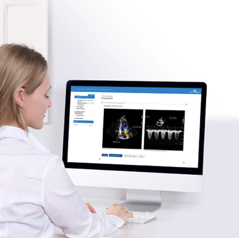
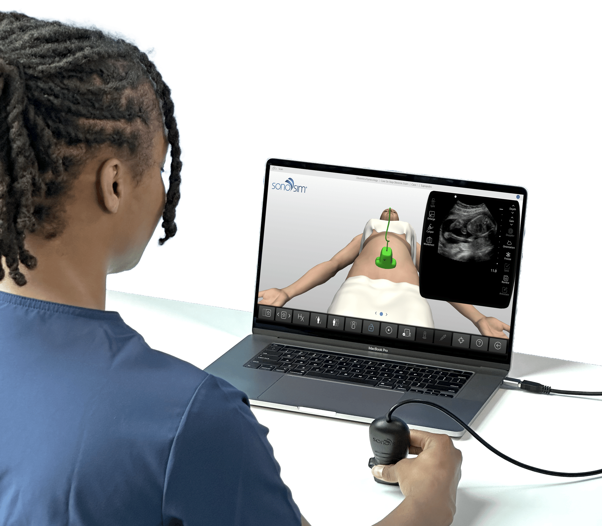
Woman completing her point of care ultrasound training with SonoSim
  • Interactive, online courses including ultrasound videos 
  • Relevant scanning cases from real patients, accessible in the SonoSimulator
  • Image acquisition & interpretation guidance, feedback, and ultrasound image portfolios
  • Automatically graded knowledge checks & A Mastery Test
  • Certificates & CME (if relevant)
Woman working on her ultrasound education in the SonoSimulator

POCUS Training for Programs, Institutions, & Practitioners

Hit Training Milestones & Standards

Our ecosystem of ultrasound training and assessment resources helps POCUS program leaders ensure providers meet existing and future POCUS ultrasound training standards. SonoSim’s ecosystem delivers each of the consensus-based knowledge elements required for achieving ultrasound competency on an application-specific basis.1

SonoSim realizes there is no substitute for bedside clinical practice & live instruction. We have created an ecosystem to supplement and support bedside clinical practice. Learners and program administrators that combine bedside clinical practice with the SonoSim ecosystem achieve the best learning outcomes. We help educators overcome the traditional barriers to ultrasound training and help learners gain the ultrasound skills needed to improve patient care. SonoSim organizes and delivers ultrasound training to deliver the following elements that have been associated with developing ultrasound competency across its training resources:

Ultrasound Competency Elements

  • Indication for the examination
  • Applied knowledge of ultrasound equipment
  • Image optimization
  • Systematic examination
  • Interpretation of images
  • Documentation of examination
  • Medical decision-making
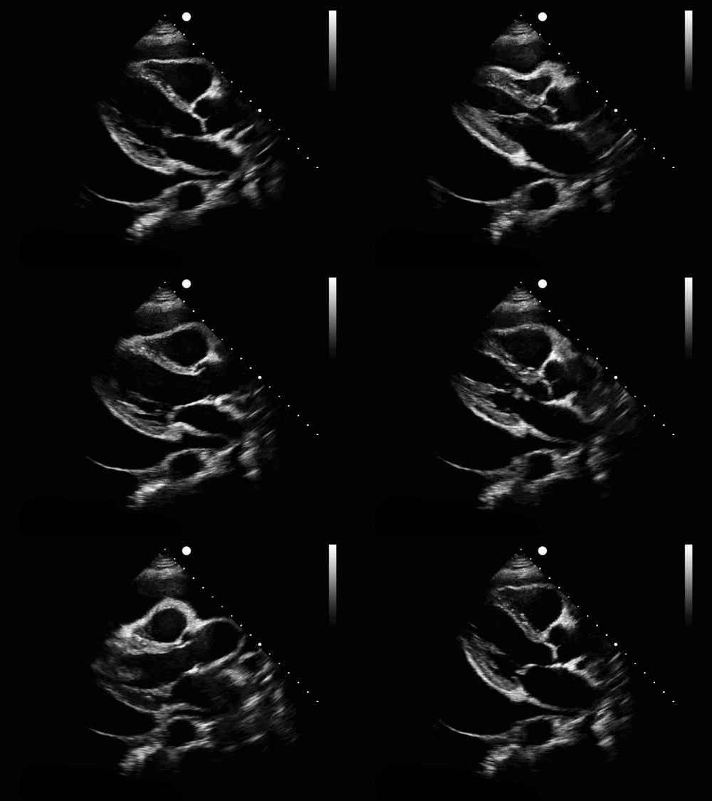
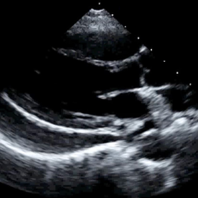
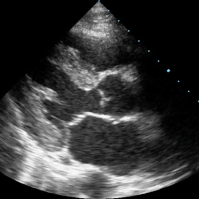
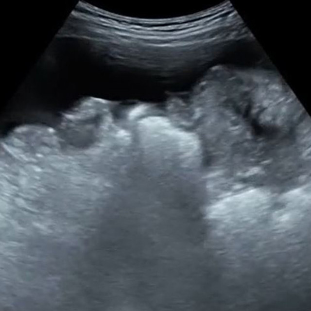
Ultrasound image of large pericardial effusion in the PLAX view

Ultrasound Training for DMS Programs

Aligned to AIUM Parameters & CAAHEP Standards

Adopted by 180+ Diagnostic Medical Sonography (DMS) programs, SonoSim transforms ordinary laptops into dynamic ultrasound training platforms. By complementing bedside clinical practice and live or remote ultrasound instruction provided by DMS programs, we help you help your learners develop ultrasound competency. Our DMS training content adheres to accreditation requirements published by the Commission on Accreditation of Allied Health Education Programs (CAAHEP), and consensus-based guidelines generated by the American Institute of Ultrasound in Medicine (AIUM).

The SonoSim Ecosystem of ultrasound training includes 80+ peer-reviewed ultrasound training topics that are tailored to develop ultrasound knowledge. Ultrasound image acquisition, interpretation, access to thousands of real pathologic cases, and adherence to DMS imaging protocols are taught using the patented laptop-based SonoSimulator®.  Study tools, like SPI Test Prep, ensure your students are optimally prepared for Board certification exams, while SonoSim’s QuestionBank allows them to engage in interactive case-based ultrasound learning. In addition, our performance tracking dashboard, automated ultrasound image review and feedback application, and curriculum integration tools simplify the process of integrating SonoSim into your curriculum. 

SonoSim ultrasound training topics cover the DMS General Education Curriculum, including SPI Test Preparation, as well as the following concentrations:
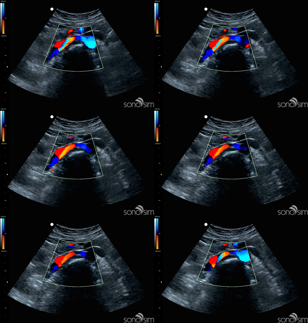
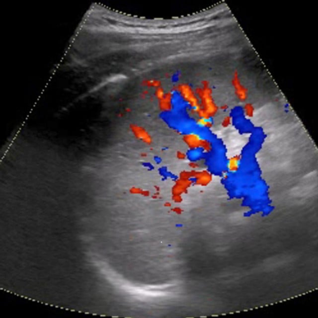
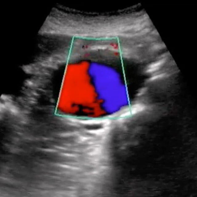
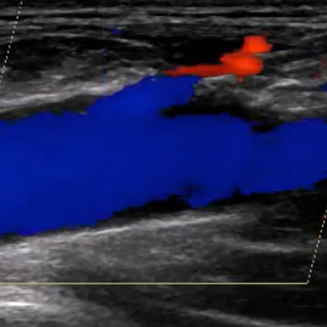
Renal arteries doppler image found in SonoSim ultrasound training cases
  • Abdomen
  • OB/GYN
  • Vascular
  • Adult Cardiac
  • Breast
  • Musculoskeletal

Ultrasound Training Topic Categories

  • Anatomy & Physiology

  • Core Clinical

  • Advanced Clinical

  • U/S-Guided Procedures

Screen Shot 2023-01-23 at 8.36.57 PM

Anatomy & Physiology Ultrasound Training

Our A&P ultrasound training provides a strong foundation of ultrasound knowledge, specific to anatomical regions, organs, and structures. Our expert-led ultrasound courses cover everything from regional anatomy & physiology to sonographic anatomy, scanning technique, and imaging tips & pitfalls. Relevant scanning cases from real patients are accessible in the SonoSimulator for each topic.
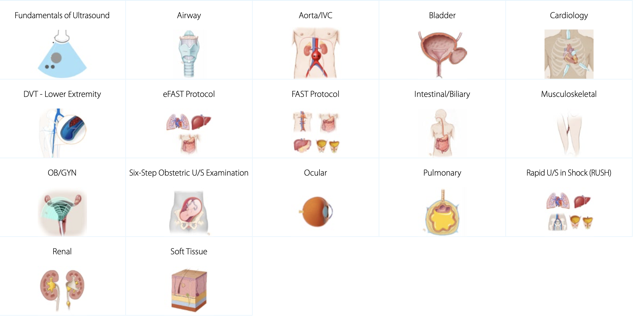
Screen Shot 2023-01-23 at 8.44.43 PM

Core Clinical Ultrasound Training

Our clinical ultrasound training topics provide in-depth comprehension of the necessary components to accurately assess and diagnose using ultrasound. We cover exam indications, regional anatomy, sonographic anatomy, and sonographic technique. Our ultrasound courses use pathologic case studies and imaging tips & pitfalls to give you an in-depth understanding of how diseases present in ultrasound imaging. Associated scanning cases from real patients are included and accessible in the SonoSimulator. This topic category supports both diagnostic medical sonography (DMS) learners as well as medical practitioners looking for point-of-care ultrasound (POCUS) training.
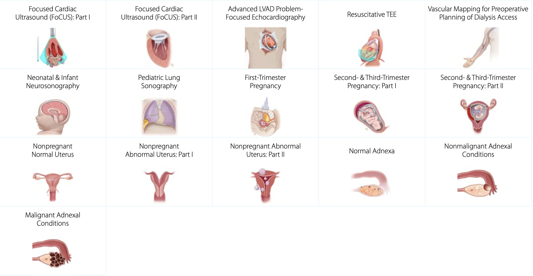
Screen Shot 2023-01-23 at 8.45.56 PM

Advanced Clinical Ultrasound Training

Our advanced clinical ultrasound topics cover complex diagnoses and sonographic applications. This is an ever-expanding list of topics with a focus on specific pathologic conditions, providing learners a deep understanding of ultrasound techniques for assessing a variety of complex medical conditions. Each topic is accompanied with 10-20 scanning cases from real patients, that are accessed in the SonoSimulator.
acute care practitioners ultrasound training

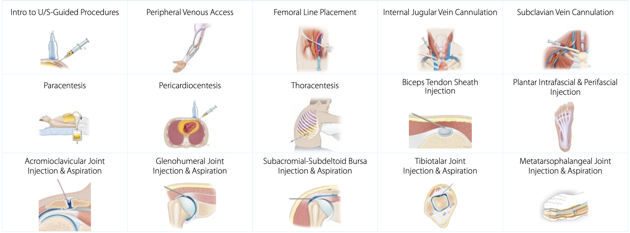
Ultrasound-Guided Procedure Training

Ultrasound-guided procedures are increasingly used to minimize complications and improve patient care. SonoSim ultrasound procedure training covers patient positioning, procedural steps, imaging adjuncts, potential complications, and more. Scanning cases from real patients are also provided with these topics, to support unlimited practice of the cognitive tasks required to perform ultrasound-guided procedures.  Practice as much as you like, risk-free - anytime, anywhere. 

SonoSim Ultrasound Training

SonoSim helps educators develop competency in students & learners, and supports healthcare providers in developing ultrasound expertise. Explore how easy & efficient ultrasound training can be with SonoSim.

SonoSim cloud-based, multimedia, didactic courses are created by leading ultrasound experts and are internet accessible immediately after purchase, on any device. There are knowledge checks with real-time feedback throughout each course, as well as a Mastery Test that is automatically graded.

The SonoSimulator® helps develop and maintain the critical visuomotor and visuospatial skills that are central to image acquisition and interpretation with real patient imagery, expert tutorials on-demand, and real-time feedback on success.

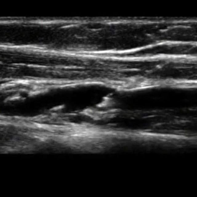
Aorta IVC ultrasound image used in point of care ultrasound training
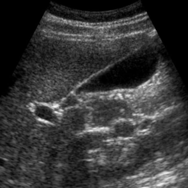
Ultrasound image of the biliary tree used in ultrasound education
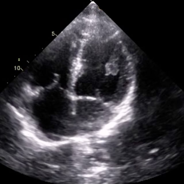
An ultrasound scan of the heart used in cardiac ultrasound training
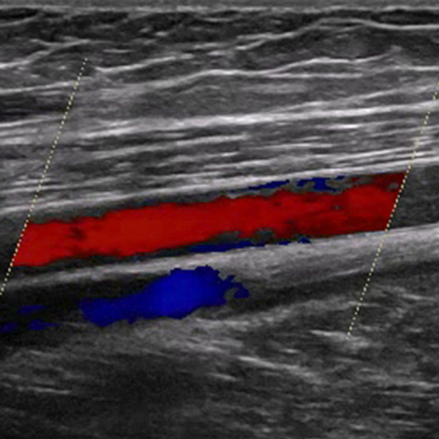
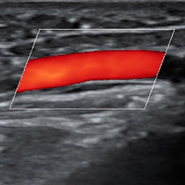
Leg arterial ultrasound education image
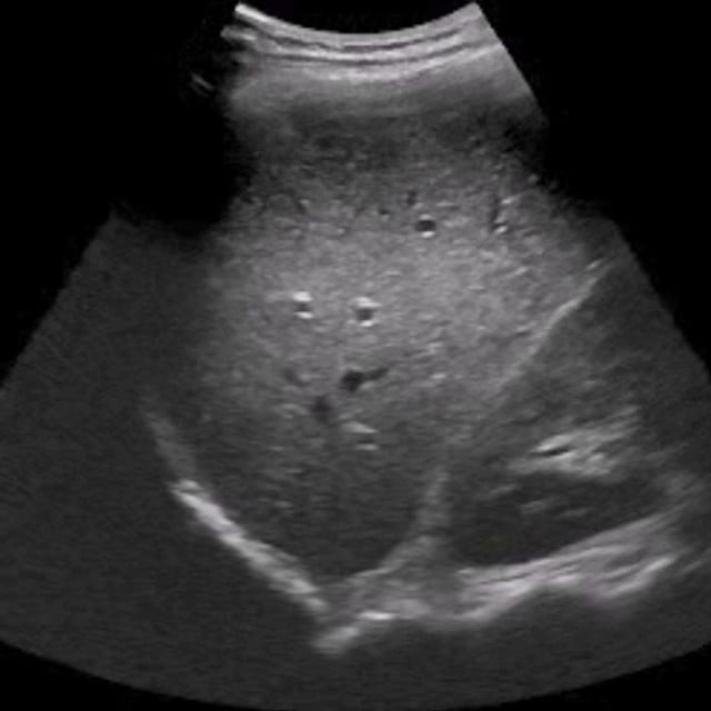
Ultrasound of the liver used in point of care ultrasound training
Ultrasound training image of the spleen

Ultrasound Physics & Fundamentals Introductory Training

Image Carrousel 74

Learn the fundamentals of using ultrasound in the clinical setting. Ultrasound physics, typical imaging parameters, artifacts, and modes are covered in detail, as well as how to operate ultrasound systems, controlling for infection and other safety considerations.

Course Highlights

SonoSimulator Scanning Cases

The course in this module covers topics including:

  • Ultrasound Instrumentation & Image Acquisition
  • Basic Ultrasound Physics
  • Transducers
  • Imaging Conventions including Transducer Orientation and Procedure Preparation
  • Imaging Modes & Artifacts
  • Doppler-Mode Imaging
  • Ultrasound System Operation
  • Infection Control and Biosafety

The 10 SonoSim SkillBox scanning case in this module include and cover:

  • 27 Interspersed Questions & Answers with Real-Time Feedback
  • 20-Question Mastery Test
  • Transducer Types: Linear, Curvilinear, Phased Array

How to buy5

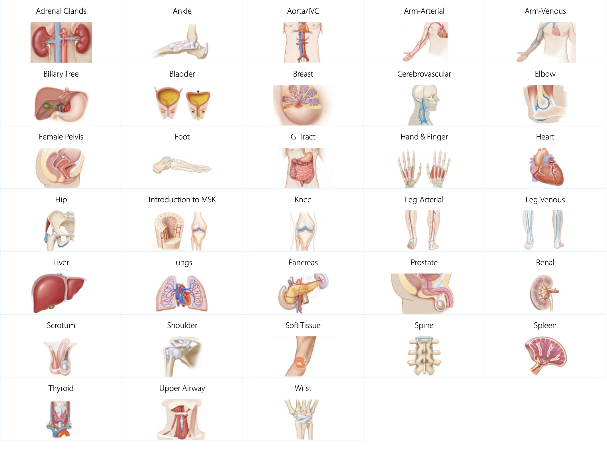
Anatomy & Physiology Series

SonoSim offers a wide array of courses that align learning sonographic anatomy & physiology with traditional textbook anatomy & physiology. Each SonoSim Anatomy & Physiology topic comes with an interactive, multimedia online course including ultrasound videos, knowledge checks and a Mastery Test, as well as a series of normal anatomy scanning cases from real patients to scan in the SonoSimulator. This content is geared toward those starting out in ultrasound or adding an anatomical area of competency not previously studied.

Adrenal Glands Anatomy & Physiology Ultrasound Training

Image Carrousel 2

Learn the relationship of traditional illustrative and sonographic anatomy of the adrenal glands and how to interpret ultrasound imaging of the region. Learn sonographic techniques and scanning approaches used in ultrasound imaging to assess the adrenals. Learn to identify and characterize the adrenal glands with ultrasonography.

Course Highlights

SonoSimulator Scanning Cases

The course in this module covers topics including:

  • Adrenal Anatomy & Physiology, including Vascular Supply
  • Sonographic Anatomy of the Adrenal Glands
  • Sonographic Techniques in Adrenal Gland Imaging
  • Pearls & Pitfalls when Evaluating the Adrenal Glands

The 3 real patient scanning cases in this module include and cover:

  • Patient Types: Adult Only, Male & Female
  • Relevant Findings/Pathology: Normal Examples of Adrenal Gland Anatomy
  • Transducer Types: Curvilinear
  • Special Case Features: Additional Abdominal CT imaging, Anatomic Layer Removal

How to buy5

Ankle Anatomy & Physiology Ultrasound Training

Image Carrousel 4

Learn the relationship of traditional illustrative and sonographic anatomy of the ankle and how to interpret ultrasound imaging of the ankle. Learn sonographic techniques and scanning approaches used in ultrasound imaging to assess the ankle. Learn to identify and characterize the ankle with ultrasonography.

Course Highlights

SonoSimulator Scanning Cases

The course in this module covers topics including:

  • Ankle Anatomy including Joint Structures
  • Physiology & Ankle Movement
  • Sonographic Anatomy of the Ankle
  • Sonographic Techniques
  • Pearls & Pitfalls when Evaluating the Ankle

The 4 real patient scanning cases in this module include and cover:

  • Patient Types: Adult Only, Male and Female
  • Relevant Findings/Pathology: Normal Examples of Ankle Anatomy
  • Transducer Types: Linear
  • Special Case Features: Anatomic Layer Removal

How to buy5


Aorta & Inferior Vena Cava ( IVC ) Anatomy & Physiology Ultrasound Training

Image Carrousel 5

Learn the relationship of traditional illustrative and sonographic anatomy of the aorta and inferior vena cava (IVC) and how to interpret ultrasound imaging of the aorta & IVC. Learn sonographic techniques and scanning approaches used in ultrasound imaging to assess the aorta and inferior vena cava. Learn to identify and characterize the aorta / IVC with ultrasonography.

Course Highlights

SonoSimulator Scanning Cases

The course in this module covers topics including:

  • Anatomy and Physiology of the Aorta and Inferior Vena Cava
  • Sonographic Anatomy of the Aorta & Inferior Vena Cava
  • Sonographic Techniques including Doppler Adjuncts
  • Pearls & Pitfalls when Evaluating the Aorta and Inferior Vena Cava

The 5 real patient scanning cases in this module include and cover:

  • Patient Types: Adult Only, Male & Female
  • Relevant Findings/Pathology: Normal Examples of Aorta and IVC Anatomy
  • Doppler & Imaging Modes: Color-Flow Doppler, Pulsed-Wave Doppler
  • Transducer Types: Curvilinear
  • Special Case Features: Additional Abdominal CT imaging, Anatomic Layer Removal

How to buy5


Arm Arterial Anatomy & Physiology Ultrasound Training

Image Carrousel 7

Learn the relationship of traditional illustrative and sonographic anatomy of the arm and how to interpret ultrasound imaging of the upper extremity arterial system. Learn sonographic techniques and scanning approaches used in ultrasound imaging of the upper extremity arterial system. Learn to identify and characterize the macroscopic and regional anatomical components of the upper extremity arterial system with ultrasonography.

Course Highlights

SonoSimulator Scanning Cases

The course in this module covers topics including:

  • Upper Extremity Arterial Anatomy
  • Arm Arterial Physiology Including Circulation & Blood Flow
  • Sonographic Anatomy of Upper Extremity Arteries
  • Sonographic Techniques
  • Pearls & Pitfalls when Evaluating the Arteries

The 5 real patient scanning cases in this module include and cover:

  • Patient Types: Adult Only, Male & Female
  • Relevant Findings/Pathology: Normal Examples of Arterial Arm Anatomy
  • Doppler & Imaging Modes: Color-Flow Doppler, Pulsed-Wave Doppler
  • Transducer Types: Linear
  • Sonographic Technique
  • Special Case Features: Compression, Anatomic Layer Removal

How to buy5


Arm Venous Anatomy & Physiology Ultrasound Training

Image Carrousel 8

Learn the relationship of traditional illustrative and sonographic anatomy of the arm and how to interpret ultrasound imaging of arm veins. Learn sonographic techniques and scanning approaches used in ultrasound imaging to assess the venous system of the upper extremities. Learn to identify and characterize arm veins with ultrasonography.

Course Highlights

SonoSimulator Scanning Cases

The course in this module covers topics including:

  • Arm Vein Anatomy including Deep and Superficial Veins
  • Venous Arm Physiology including Capacity and Flow
  • Sonographic Anatomy of the Arm Veins
  • Sonographic Anatomy
  • Sonographic Techniques
  • Pearls & Pitfalls when Evaluating the Arm Veins

The 5 real patient scanning cases in this module include and cover:

  • Patient Types: Adult Only, Male & Female
  • Relevant Findings/Pathology: Normal Examples of Venous Arm Anatomy
  • Doppler & Imaging Modes: Color-Flow Doppler, Pulsed-Wave Doppler
  • Transducer Types: Linear
  • Special Case Features: Compression, Anatomic Layer Removal

How to buy5


Biliary Tree Anatomy & Physiology Ultrasound Training

Image Carrousel 11

Learn the relationship of traditional illustrative and sonographic anatomy of the biliary tree and how to interpret ultrasound imaging of the gallbladder, vascular supply and surrounding structures. Learn sonographic techniques and scanning approaches used in ultrasound imaging to assess the biliary tree including bile storage and enterohepatic circulation. Learn to identify and characterize the macroscopic and regional anatomical components of the biliary tract with ultrasonography.

Course Highlights

SonoSimulator Scanning Cases

The course in this module covers topics including:
  • Biliary Tract & Gallbladder Anatomy, including Bile Ducts & Neurovascular Supply
  • Gallbladder & Biliary Tract Physiology, including Bile Storage and Enterohepatic Circulation
  • Sonographic Anatomy of the Biliary Tree, including differentiation between Intestines and Aorta and Inferior Vena Cava
  • Sonographic Anatomy
  • Sonographic Techniques
  • Pearls & Pitfalls when Evaluating the Biliary Tree
The 3 real patient scanning cases in this module include and cover:
  • Patient Types: Male & Female, 2 Adult & 1 Pediatric
  • Relevant Findings/Pathology: Normal Examples of Biliary Tree Anatomy
  • Transducer Types: Curvilinear
  • Special Case Features: Additional Abdominal CT Imaging

How to buy5


Bladder Anatomy & Physiology Ultrasound Training

Image Carrousel 12

Learn the relationship of traditional illustrative and sonographic anatomy of the bladder and how to interpret ultrasound imaging of the bladder and surrounding structures. Learn sonographic techniques and scanning approaches used in ultrasound imaging to assess the bladder including bladder volume measurements. Learn to identify and characterize the bladder with ultrasonography.

Course Highlights

SonoSimulator Scanning Cases

The course in this module covers topics including:

  • Bladder Anatomy on the Axial, Coronal, & Sagittal Plane
  • Physiology of the Bladder
  • Sonographic Anatomy of the Pelvis, including Female-specific Variation
  • Sonographic Techniques, including Male and Female Variables
  • Sonographic Technique
  • Bladder Volume Measurements
  • Pearls & Pitfalls when Evaluating the Bladder

The 5 real patient scanning cases in this module include and cover:

  • Patient Types: Male & Female, 4 Adult & 1 Pediatric
  • Relevant Findings/Pathology: Normal Examples of Bladder Anatomy
  • Doppler & Imaging Modes: Color-Flow Doppler
  • Transducer Types: Curvilinear
  • Special Case Features: Additional Abdominal/Pelvic CT Imaging, Anatomic Layer Removal

How to buy5


Breast Anatomy & Physiology Ultrasound Training

Image Carrousel 14

Learn the relationship of traditional illustrative and sonographic anatomy and how to interpret ultrasound imaging of the breast. Learn sonographic techniques and scanning approaches used in ultrasound imaging to assess the breast including breast layers, normal anatomical variants and age-related changes. Learn to identify and characterize the breast with ultrasonography.

Course Highlights

SonoSimulator Scanning Cases

The course in this module covers topics including:

  • Breast Anatomy & Physiology
  • Sonographic Anatomy of the Breast
  • Discerning Breast Layers, Normal Anatomical Variants, & Age-Related Changes
  • Sonographic Breast Scanning Techniques
  • Pearls & Pitfalls when Evaluating the Breast

The 3 real patient scanning cases in this module include and cover:

  • Patient Types: Adult Only, Female
  • Relevant Findings/Pathology: Normal Examples of Breast Anatomy
  • Transducer Types: Linear
  • Special Case Features: Additional Breast CT and MRI Imaging, Anatomic Layer Removal

How to buy5


Cerebrovascular Anatomy & Physiology Ultrasound Training

Image Carrousel 16

Learn the relationship of traditional illustrative and sonographic anatomy of the cerebral vasculature and how to interpret cerebrovascular ultrasound imaging. Learn sonographic techniques and scanning approaches used in ultrasound imaging to assess cerebral vasculature. Learn to identify and characterize cerebral vasculature with ultrasonography.

Course Highlights

SonoSimulator Scanning Cases

The course in this module covers topics including:

  • Cerebral Blood Supply
  • Cerebrovascular Physiology
  • Histophysiology
  • Cerebrovascular Sonographic Anatomy
  • Sonographic Techniques for scanning Carotid & Vertebral Arteries & the Internal Jugular Vein
  • Pearls & Pitfalls when Evaluating the Cerebrovascular System

The 5 real patient scanning cases in this module include and cover:

  • Patient Types: Adult Only, Male & Female
  • Relevant Findings/Pathology: Normal Examples of Cerebrovascular Anatomy
  • Doppler & Imaging Modes: Color-Flow Doppler, Pulsed-Wave Doppler
  • Transducer Types: Linear
  • Special Case Features: Additional Cerebrovascular MRA Imaging, Anatomic Layer Removal

How to buy5


Elbow Anatomy & Physiology Ultrasound Training

Image Carrousel 19

Learn the relationship of traditional illustrative and sonographic anatomy and how to interpret ultrasound imaging of the elbow. Learn sonographic techniques and scanning approaches used in ultrasound imaging to assess the elbow. Learn to identify and characterize the elbow with ultrasonography.

Course Highlights

SonoSimulator Scanning Cases

The course in this module covers topics including:

  • Elbow Anatomy, including Joint Structures and Musculature
  • Elbow Movement / Elbow Physiology
  • Sonographic Anatomy of the Four (4) Quadrants of the Elbow
  • Sonographic Techniques
  • Pearls & Pitfalls when Evaluating the Elbow

The 4 real patient scanning cases in this module include and cover:

  • Patient Types: Adult Only, Male
  • Relevant Findings/Pathology: Normal Examples of Elbow Anatomy
  • Transducer Types: Linear

How to buy5


Female Pelvis Anatomy & Physiology Ultrasound Training

Image Carrousel 21

Learn the relationship of traditional illustrative and sonographic anatomy and how to interpret ultrasound imaging of the female pelvis. Learn sonographic techniques and scanning approaches used in ultrasound imaging to assess the female pelvis. Learn to identify and characterize the female pelvis with ultrasonography.

Course Highlights

SonoSimulator Scanning Cases

The course in this module covers topics including:

  • Female Pelvis Anatomy, including The Reproductive Tract
  • Female Pelvis Physiology, including Histophysiology & Embryology
  • Sonographic Anatomy & Techniques, including Transabdominal and Endovaginal Imaging
  • Pearls & Pitfalls when Evaluating the Female Pelvis

The 3 real patient scanning cases in this module include and cover:

  • Patient Types: Female, 2 Adult & 1 Pediatric
  • Relevant Findings/Pathology: Normal Examples of Female Pelvic Anatomy
  • Transducer Types: Curvilinear
  • Special Case Features: Anatomic Layer Removal

How to buy5


Foot Anatomy & Physiology Ultrasound Training

Image Carrousel 26

Learn the relationship of traditional illustrative and sonographic anatomy and how to interpret ultrasound imaging of the foot. Learn sonographic techniques and scanning approaches used in ultrasound imaging to assess the foot. Learn to identify and characterize the foot with ultrasonography.

Course Highlights

SonoSimulator Scanning Cases

The course in this module covers topics including:

  • Anatomy & Physiology of the Foot, including Bones, Joints & Musculature of the Foot
  • Neurovascular Supply to the Foot
  • Sonographic Imaging Techniques for the Dorsal and Plantar Foot
  • Pearls & Pitfalls in Evaluating the Foot

The 3 real patient scanning cases in this module include and cover:

  • Patient Types: Adult Only, Male & Female
  • Relevant Findings/Pathology: Normal Examples of Foot Anatomy
  • Transducer Types: Linear
  • Special Case Features: Additional Foot MRI Imaging

How to buy5


GI Tract Anatomy & Physiology Ultrasound Training

Image Carrousel 27

Learn the relationship of traditional illustrative and sonographic anatomy and how to interpret ultrasound imaging of the gastrointestinal tract, including the mouth, pharynx, esophagus, stomach, intestines, and colon. Learn sonographic techniques and scanning approaches used in ultrasound imaging to assess the GI tract. Learn to identify and characterize the gastrointestinal system using ultrasonography.

Course Highlights

SonoSimulator Scanning Cases

The course in this module covers topics including:

  • Gastrointestinal Anatomy of the Upper and Lower GI Tracts
  • GI Tract Physiology including Mouth, Pharynx, Esophagus, Intestinal Digestion, Absorption, Intestinal Peristalsis, and Colorectal Function
  • Sonography of the Complete Gastrointestinal ( GI ) Tract
  • Sonographic Techniques
  • Pearls & Pitfalls when Evaluating the GI Tract

The 3 real patient scanning cases in this module include and cover:

  • Patient Types: Adult Only, Male & Female
  • Relevant Findings/Pathology: Normal Examples of GI Tract Anatomy
  • Transducer Types: Linear, Curvilinear
  • Special Case Features: Compression, Anatomic Layer Removal

How to buy5


Hand & Finger Anatomy & Physiology Ultrasound Training

Image Carrousel 35

Learn the relationship of traditional illustrative and sonographic anatomy of the hand and fingers and how to interpret ultrasound imaging of the hand and fingers. Learn sonographic techniques and scanning approaches used in ultrasound imaging to assess the hand and fingers. Learn to identify and characterize the hand and fingers with ultrasonography.

Course Highlights

SonoSimulator Scanning Cases

The course in this module covers topics including:

  • Hand & Finger Anatomy & Physiology including Kinematics, Muscles, Tendons, & Ligaments
  • Neurovascular Supply to the Hand & Fingers
  • Sonographic Anatomy of Hands & Fingers
  • Sonographic Techniques for Fingers, Palms Tendons & Ligaments
  • Pearls & Pitfalls when Evaluating the Hand & Fingers

The 5 real patient scanning cases in this module include and cover:

  • Patient Types: Adult Only, Male
  • Relevant Findings/Pathology: Normal Examples of Hand & finger Anatomy
  • Transducer Types: Linear
  • Special Case Features: Anatomic Layer Removal

How to buy5


Heart Anatomy & Physiology Ultrasound Training

Image Carrousel 36

Learn the relationship of traditional illustrative and sonographic anatomy and how to interpret ultrasound imaging of the heart. Learn sonographic techniques and scanning approaches used in cardiac ultrasound imaging. Learn characterize the heart using ultrasonography.

Course Highlights

SonoSimulator Scanning Cases

The course in this module covers topics including:

  • External & Internal Heart Anatomy
  • Histophysiology
  • Cardiac Cycle
  • Circulation
  • Sonographic Anatomy of the Heart, including Parasternal Long-Axis and Four-Chamber Views
  • Sonographic Techniques, including Acquiring the Parasternal, Apical & Subcostal Windows
  • Diagnostic & Clinical Application
  • Pearls & Pitfalls when Evaluating the Heart

The 5 real patient scanning cases in this module include and cover:

  • Patient Types: Male & Female, 4 Adult & 1 Pediatric
  • Relevant Findings/Pathology: Normal examples of heart anatomy
  • Transducer Types: Phased Array
  • Special Case Features: Additional Chest CT Imaging, Anatomic Layer Removal

How to buy5


Hip Anatomy & Physiology Ultrasound Training

Image Carrousel 37

Learn the relationship of traditional illustrative and sonographic anatomy of the hip and how to interpret ultrasound imaging of the hip. Learn sonographic techniques and scanning approaches used in ultrasound imaging to assess the hip. Learn to identify and characterize the hip with ultrasonography.

Course Highlights

SonoSimulator Scanning Cases

The course in this module covers topics including:

  • Hip Anatomy & Physiology, including Joint Structures, Musculature and Neurovascular Supply
  • Sonographic Anatomy of the Anterior, Medial, Lateral & Posterior Hip
  • Sonographic Techniques, including Anterior, Medial, Lateral & Posterior Approaches
  • Pearls & Pitfalls when Evaluating the Hip

The 4 real patient scanning cases in this module include and cover:

  • Patient Types: Adult Only, Male
  • Relevant Findings/Pathology: Normal Examples of Hip Anatomy
  • Transducer Types: Linear
  • Special Case Features: Compression

How to buy5


Knee Anatomy & Physiology Ultrasound Training

Image Carrousel 40.1

Learn how to use ultrasound in the evaluation of the knee, including knee anatomy and physiology, and sonographic anatomy of the knee. Optimal transducer selection, patient positioning, imaging approaches, and techniques for sonographically evaluating the knee are discussed.

Course Highlights

SonoSimulator Scanning Cases

The course in this module covers topics including:

  • Anatomy of the Knee, including Ligaments, Joint Structures, Tendons, Muscles & Neurovascular Supply
  • Knee Kinematics
  • Sonographic Anatomy, including Anterior, Medial, Lateral & Posterior Knee Anatomy
  • Sonographic Techniques
  • Pearls & Pitfalls of Evaluating the Knee

The 4 real patient scanning cases in this module include and cover:

  • Patient Types: Adult Only, Male
  • Relevant Findings/Pathology: Normal Examples of Knee Anatomy
  • Transducer Types: Linear
  • Special Case Features: Compression

How to buy5


Leg Arterial Anatomy & Physiology Ultrasound Training

Image Carrousel 41

Learn how to use ultrasound in the evaluation of the lower extremity arterial system, including lower extremity arterial anatomy and physiology and sonographic anatomy of the lower extremity arterial system. Optimal transducer selection, patient positioning, imaging approaches, and techniques for sonographically evaluating the lower extremity arterial system are discussed.

Course Highlights

SonoSimulator Scanning Cases

The course in this module covers topics including:

  • Arterial Anatomy & Physiology of the Leg
  • Sonographic Anatomy of Leg Arteries including Doppler Adjuncts
  • Sonographic Imaging Techniques of the Lower Extremities
  • Pearls & Pitfalls when Evaluating the Leg Arteries

The course in this module covers topics including:

  • Arterial Anatomy & Physiology of the Leg
  • Sonographic Anatomy of Leg Arteries including Doppler Adjuncts
  • Sonographic Imaging Techniques of the Lower Extremities
  • Pearls & Pitfalls when Evaluating the Leg Arteries

How to buy5


Leg Venous Anatomy & Physiology Ultrasound Training

Image Carrousel 42

Learn how to use ultrasound in the evaluation of the lower extremity vasculature, including lower extremity venous anatomy and physiology, and sonographic anatomy of the lower extremity venous system. Optimal transducer selection, patient positioning, imaging approaches, and techniques for sonographically evaluating the lower extremity venous system are discussed.

Course Highlights

SonoSimulator Scanning Cases

The course in this module covers topics including:

  • Anatomy of the Venous System of the Leg including Deep and Superficial Veins
  • Venous Leg Physiology including Capacity and Flow
  • Sonographic Anatomy of Leg Veins
  • Sonographic Imaging Techniques for Hip, Thigh, Calf, Ankle & Foot Veins
  • Pearls & Pitfalls when Evaluating Leg Veins

The 5 real patient scanning cases in this module include and cover:

  • Patient Types: Male & Female, 4 Adult & 1 Pediatric
  • Relevant Findings/Pathology: Normal Examples of Venous Leg Anatomy
  • Doppler & Imaging Modes: Color-Flow Doppler, Pulsed-Wave Doppler
  • Transducer Types: Linear, Curvilinear
  • Special Case Features: Compression, Anatomic Layer Removal

How to buy5


Liver Anatomy & Physiology Ultrasound Training

Image Carrousel 43

Learn how to use ultrasound in the evaluation of the liver, including anatomy and physiology, and sonographic anatomy of the liver. Optimal transducer selection, patient positioning, imaging approaches, and techniques for sonographically evaluating the liver are discussed.

Course Highlights

SonoSimulator Scanning Cases

The course in this module covers topics including:

  • Liver Anatomy including Portal Triad & Porta Hepatis
  • Liver Physiology
  • Sonographic Anatomy of the Liver, including the Various Lobes and Planes
  • Sonographic Techniques, including the Use of Doppler Mode
  • Pearls & Pitfalls when Evaluating the Liver

The 3 real patient scanning cases in this module include and cover:

  • Patient Types: Male & Female, 2 Adult & 1 Pediatric
  • Relevant Findings/Pathology: Normal Examples of Liver Anatomy
  • Transducer Types: Curvilinear
  • Special Case Features: Additional Abdominal CT Imaging, Anatomic Layer Removal

How to buy5


Lungs Anatomy & Physiology Ultrasound Training

Image Carrousel 44

Learn how to use ultrasound in the evaluation of the lungs, including lung anatomy and physiology, and sonographic anatomy of the lungs. Optimal transducer selection, patient positioning, imaging approaches, and techniques for sonographically evaluating the lungs are discussed.

Course Highlights

SonoSimulator Scanning Cases

The course in this module covers topics including:

  • Normal Lung Anatomy including Cross-Sectional Views
  • Functional Respiration & Pulmonary Blood Flow
  • Sonographic Anatomy of the Lungs
  • Sonographic Techniques for Lung Imaging
  • Pearls & Pitfalls when Evaluating the Lung

The 5 real patient scanning cases in this module include and cover:

  • Patient Types: Male & Female, 4 Adult & 1 Pediatric
  • Relevant Findings/Pathology: Normal Examples of Lung Anatomy
  • Doppler & Imaging Modes: Color-Flow Doppler, Power Doppler, M-Mode
  • Transducer Types: Linear, Curvilinear
  • Special Case Features: Anatomic Layer Removal

How to buy5


MSK Anatomy & Physiology Introductory Ultrasound Training

Image Carrousel 47

Learn the relationship of traditional illustrative and sonographic anatomy of the musculoskeletal system, including how to interpret ultrasound imaging of the musculoskeletal system. Learn sonographic techniques and scanning approaches used in ultrasound imaging to assess the musculoskeletal system. Learn to identify and characterize the components of the musculoskeletal system with ultrasonography.

Course Highlights

SonoSimulator Scanning Cases

The course in this module covers topics including:

  • Anatomy & Physiology of Hard and Soft Tissues in the Musculoskeletal System
  • Sonographic Anatomy of Bones, Tendons, Muscles & Joints
  • Sonographic Techniques
  • Pearls & Pitfalls when Evaluating Musculoskeletal Structures

The 4 real patient scanning cases in this module include and cover:

  • Patient Types: Adult Only, Male
  • Relevant Findings/Pathology: Normal Examples of Musculoskeletal (MSK) Anatomy - Shoulder, Hand, Finger, Spine & Elbow
  • Transducer Types: Linear
  • Special Case Features: Additional MSK MRI Imaging

How to buy5


Pancreas Anatomy & Physiology Ultrasound Training

Image Carrousel 52

Learn how to use ultrasound to evaluate the pancreas, including pancreas anatomy and physiology and sonographic anatomy of the pancreas. Optimal transducer selection, patient positioning, imaging approaches, and techniques for sonographically evaluating the pancreas are discussed.

Course Highlights

SonoSimulator Scanning Cases

The course in this module covers topics including:

  • Pancreatic Anatomy & Physiology, including Vascular Supply
  • Sonographic Anatomy of the Pancreas
  • Sonographic Techniques including the Use of Doppler Imaging
  • Pearls & Pitfalls when Evaluating the Pancreas

The 3 real patient scanning cases in this module include and cover:

  • Patient Types: Male and Female, 2 Adult and 1 Pediatric
  • Relevant Findings/Pathology: Normal Examples of Pancreatic Anatomy
  • Transducer Types: Curvilinear
  • Special Case Features: Additional Abdominal CT imaging, Anatomic Layer Removal

How to buy5


Prostate Anatomy & Physiology Ultrasound Training

Image Carrousel 56

Learn how to use ultrasound in the evaluation of the prostate, including prostate anatomy and physiology, and sonographic anatomy of the prostate. Optimal transducer selection, patient positioning, imaging approaches, and techniques for sonographically evaluating the prostate are discussed.

Course Highlights

SonoSimulator Scanning Cases

The course in this module covers topics including:

  • Prostate Gland Anatomy, including Neurovascular Supply
  • Prostate Physiology
  • Sonographic Anatomy of the Prostate
  • Sonographic Techniques for Prostate Imaging
  • Pearls & Pitfalls when Evaluating the Prostate

The 3 real patient scanning cases in this module include and cover:

  • Patient Types: Adult Only, Male
  • Relevant Findings/Pathology: Normal Examples of Prostate Anatomy
  • Transducer Types: Curvilinear
  • Special Case Features: Additional Prostate MRI Imaging, Anatomic Layer Removal

How to buy5

Renal Anatomy & Physiology Ultrasound Training

Image Carrousel 58

Learn how to use ultrasound in the evaluation of the kidneys, including kidney anatomy and physiology and sonographic anatomy of the kidneys. Optimal transducer selection, patient positioning, imaging approaches, and techniques for sonographically evaluating the kidneys are discussed.

Course Highlights

SonoSimulator Scanning Cases

The course in this module covers topics including:

  • Anatomy of the Kidneys Ureters & Bladder, including Renal Imaging Planes
  • Renal Physiology including Vasculature & Arterial Supply
  • Sonographic Anatomy of the Kidney & Bladder
  • Sonographic Techniques for Evaluating Both Kidneys, analyzing Renal Blood Flow, and Evaluating the Bladder

The 5 real patient scanning cases in this module include and cover:

  • Patient Types: Adult Only, Male & Female
  • Relevant Findings/Pathology: Normal Examples of Renal Anatomy
  • Doppler & Imaging Modes: Color-Flow Doppler, Pulsed-Wave Doppler
  • Transducer Types: Curvilinear
  • Special Case Features: Anatomic Layer Removal

How to buy5


Scrotum Anatomy & Physiology Ultrasound Training

Image Carrousel 62

Learn how to use ultrasound in the evaluation of the scrotum, including scrotum anatomy and physiology and sonographic anatomy of the scrotum. Optimal transducer selection, patient positioning, imaging approaches, and techniques for sonographically evaluating the scrotum are discussed.

Course Highlights

SonoSimulator Scanning Cases

The course in this module covers topics including:

  • Scrotum Anatomy & Physiology
  • Sonographic Technique & Scanning Approaches
  • Neurovascular Supply
  • Pearls & Pitfalls when Evaluating the Scrotum

The 3 real patient scanning cases in this module include and cover:

  • Patient Types: Adult Only, Male
  • Relevant Findings/Pathology: Normal Examples of Scrotal Anatomy
  • Transducer Types: Linear

How to buy5


Shoulder Anatomy & Physiology Ultrasound Training

Image Carrousel 65

Learn to use ultrasound in the evaluation of the shoulder, including shoulder anatomy and physiology, and sonographic anatomy of the shoulder. Optimal transducer selection, patient positioning, imaging approaches, and techniques for sonographically evaluating the shoulder are discussed.

Course Highlights

SonoSimulator Scanning Cases

The course in this module covers topics including:

  • Shoulder Anatomy & Physiology including Bones, Joints, Musculature, Tendons, Bursae and Neurovascular Supply
  • Sonographic Anatomy of the Shoulder
  • Sonographic Techniques for the Shoulder
  • Pearls & Pitfalls when Evaluating the Shoulder

The 3 real patient scanning cases in this module include and cover:

  • Patient Types: Adult Only, Male & Female
  • Relevant Findings/Pathology: Normal Examples of Shoulder Anatomy
  • Transducer Types: Linear

How to buy5


Soft Tissue Anatomy & Physiology Ultrasound Training

Image Carrousel 66

Learn how to use ultrasound in the evaluation of the soft tissues, including soft tissue anatomy and physiology and sonographic anatomy of soft tissues. Optimal transducer selection, patient positioning, imaging approaches, and techniques for sonographically evaluating soft tissue are discussed.

Course Highlights

SonoSimulator Scanning Cases

The course in this module covers topics including:

  • Soft Tissue Anatomy & Physiology including Histophysiology, Muscles, Tendons, Ligaments, Blood Vessels & Nerves
  • Sonographic Anatomy, including Differentiation of Bone vs. Soft Tissue
  • Sonographic Techniques, including Doppler Adjunct

The 4 real patient scanning cases in this module include and cover:

  • Patient Types: Adult Only, Male
  • Relevant Findings/Pathology: Normal Examples of Soft Tissue Anatomy
  • Transducer Types: Linear
  • Special Case Features: Additional Shoulder & Wrist MRI Imaging

How to buy5


Spine Anatomy & Physiology Ultrasound Training

Image Carrousel 68

Learn how to use ultrasound in the evaluation of the spine, including spine anatomy and physiology, and sonographic anatomy of the spine. Optimal transducer selection, patient positioning, imaging approaches, and techniques for sonographically evaluating the spine are discussed.

Course Highlights

SonoSimulator Scanning Cases

The course in this module covers topics including:

  • Spine Anatomy including Vertebra, Musculature and Spinal Cord
  • Physiology of the Spine
  • Sonographic Anatomy of Vertebral Column
  • Sonographic Techniques for Spine Imaging
  • Pearls & Pitfalls when Evaluating the Spine

The 3 real patient scanning cases in this module include and cover:

  • Patient Types: Adult Only, Male
  • Relevant Findings/Pathology: Normal Examples of Spinal Anatomy
  • Transducer Types: Curvilinear

How to buy5


Spleen Anatomy & Physiology Ultrasound Training

Image Carrousel 69

Learn how to use ultrasound in the evaluation of the spleen, including spleen anatomy and physiology, and sonographic anatomy of the spleen. Optimal transducer selection, patient positioning, imaging approaches, and techniques for sonographically evaluating the spleen are discussed.

Course Highlights

SonoSimulator Scanning Cases

The course in this module covers topics including:

  • Anatomy & Physiology of the Spleen
  • Sonographic Anatomy of the Spleen and Associated Vasculature
  • Sonographic Techniques for Spleen Imaging
  • Pearls & Pitfalls when Evaluating the Spleen

The 3 real patient scanning cases in this module include and cover:

  • Patient Types: Adult Only, Male & Female
  • Relevant Findings/Pathology: Normal Examples of Splenic Anatomy
  • Transducer Types: Curvilinear
  • Special Case Features: Additional Abdominal CT Imaging, Anatomic Layer Removal

How to buy5


Thyroid Anatomy & Physiology Ultrasound Training

Image Carrousel 72

Learn how to use ultrasound in the evaluation of the thyroid, including thyroid anatomy and physiology, and sonographic anatomy of the thyroid. Optimal transducer selection, patient positioning, imaging approaches, and techniques for sonographically evaluating the thyroid are discussed.

Course Highlights

SonoSimulator Scanning Cases

The course in this module covers topics including:

  • Anatomy & Physiology of the Thyroid & Parathyroid
  • Calcium Homeostasis
  • Sonographic Anatomy of the Thyroid & Adjacent Structures
  • Sonographic Techniques in Thyroid Imaging
  • Pearls & Pitfalls when Evaluating the Thyroid

The 5 real patient scanning cases in this module include and cover:

  • Patient Types: Adult Only, Male & Female
  • Relevant Findings/Pathology: Normal Examples of Thyroid Anatomy
  • Doppler & Imaging Modes: Color-Flow Doppler
  • Transducer Types: Linear
  • Special Case Features: Additional Neck CT Imaging, Anatomic Layer Removal

How to buy5


Upper Airway Anatomy & Physiology Ultrasound Training

Image Carrousel 79

Learn how to use ultrasound in the evaluation of the upper airway, including upper airway anatomy and physiology, and sonographic anatomy of the upper airway. Optimal transducer selection, patient positioning, imaging approaches, and techniques for sonographically evaluating the upper airway are discussed.

Course Highlights

SonoSimulator Scanning Cases

The course in this module covers topics including:

  • Anatomy of the Upper Airway including the Larynx, Hyoid Bone, Vocal Cords, Pharyngeal Elements & More
  • Upper Airway Physiology including Deglutition and Phonation
  • Sonographic Anatomy of the Upper Airway, including the Epiglottis, Trachea & Esophagus
  • Sonographic Techniques in Imaging the Throat Area
  • Pearls & Pitfalls when Evaluating the Upper Airway

The 5 real patient scanning cases in this module include and cover:

  • Patient Types: Male & Female. 4 Adult & 1 Pediatric
  • Relevant Findings/Pathology: Normal Examples of Upper Airway Anatomy
  • Doppler & Imaging Modes: Color-Flow Doppler
  • Transducer Types: Linear
  • Special Case Features: Additional Neck CT Imaging, Anatomic Layer Removal

How to buy5


Wrist Anatomy & Physiology Ultrasound Training

Image Carrousel 81

Learn how to use ultrasound in the evaluation of the wrist, including wrist anatomy and physiology, and sonographic anatomy of the wrist. Optimal transducer selection, patient positioning, imaging approaches, and techniques for sonographically evaluating the wrist are discussed.

Course Highlights

SonoSimulator Scanning Cases

The course in this module covers topics including:

  • Anatomy of the Dorsal and Volar Wrist
  • Physiology of the Wrist Joints
  • Sonographic Anatomy of the Wrist
  • Sonographic Techniques & Considerations when Imaging the Wrist, including Dynamic Scanning Techniques and the Use of Doppler-Mode Imaging
  • Pearls & Pitfalls when Evaluating the Wrist

The SonoSimulator® helps develop and maintain the critical visuomotor and visuospatial skills that are central to image acquisition and interpretation with real patient imagery, expert tutorials on-demand, and real-time feedback on success.

The 3 real patient scanning cases in this module include and cover:

  • Patient Types: Adult Only, Male
  • Relevant Findings/Pathology: Normal Examples of Wrist Anatomy
  • Transducer Types: Linear
  • Special Case Features: Additional Wrist MRI Imaging

How to buy5


Can I change my plan later?

Lorem ipsum dolor sit amet, consectetur adipiscing elit, sed do eiusmod tempor incididunt ut labore et dolore magna aliqua. Ut enim ad minim veniam, quis nostrud exercitation ullamco laboris nisi ut aliquip ex ea commodo consequat. Duis aute irure dolor in reprehenderit in voluptate velit esse cillum dolore eu fugiat nulla pariatur.

Clinical aorta IVC ultrasound education image
Clinical cardiology ultrasound training scan
Clinical deep-vein thrombosis ultrasound education scanscan
Clinical MSK ultrasound training image
Clinical RUSH protocol ultrasound image for point of care ultrasound training
Clinical soft tissue ultrasound image

Core Clinical Series

This Clinical Series is excellent for providing DMS learners with access to learn on real pathologic cases. It also covers all the recommended "Core Ultrasound Applications" recommended for POCUS practitioners by professional medical societies.

SonoSim offers an expansive library of clinical ultrasound topics to choose from. This content is geared toward intermediate learners who already have a strong understanding of sonographic anatomy in a particular region. Each SonoSim Clinical Ultrasound topic comes with an interactive, multimedia online course including ultrasound videos, knowledge checks and a Mastery Test, as well as a series of normal & pathologic scanning cases from real patients to scan in the SonoSimulator. In addition, each topic comes with CME*. This content is geared toward those looking to add an area of core clinical competency to their ultrasound skills.

Airway Clinical Ultrasound Training

Image Carrousel 3.1

Learn the basic principles and physics of point-of-care airway ultrasound imaging and how to optimize image acquisition of the human airway. Learn to evaluate the airway in an emergency and direct mainstem intubation. Learn how to use ultrasound in the evaluation of the emergent airway to confirm tracheal intubation.

Course Highlights

SonoSimulator Scanning Cases

The course in this module covers topics including:

  • Ultrasound Applications
  • Airway Anatomy
  • Sonography Anatomy & Characteristics
  • Transducer Selection & Settings
  • Ergonomic Considerations
  • Patient Positioning
  • Infection Prevention
  • Survey Scan & Procedure Steps
  • Doppler-Mode & M-Mode Imaging
  • Tips, Pitfalls, & Complications

The 10 real patient scanning cases in this module include and cover:

  • Patient Types: Male and Female, 8 Adult & 2 Pediatric
  • Relevant Findings/Pathology: Normal Adult & Pediatric Patients, Obese Patient Upper Airway Case, Endotracheal Intubation Patients, Esophageal Intubation Case, Mainstem Bronchus Intubation Case, Endotracheal Tube Localization Case, Blunt Neck Trauma Patient
  • Scanning Approaches: Vocal Cord Imaging, Esophagus Imaging, Lung Imaging, Hemidiaphragm Imaging, Detection of Intubation, Tracheal Intubation, Endotracheal Intubation
  • Transducer Types: Linear, Curvilinear, Phased Array
  • Special Case Features: Anatomic Layer Removal

How to buy5


Aorta & Inferior Vena Cava ( IVC ) Clinical Ultrasound Training

Image Carrousel 6

Learn how to evaluate aortic anatomy and identify abdominal aortic aneurysms (AAAs) with ultrasound. How to evaluate IVC anatomy and how to estimate central venous pressure (CVP) are also discussed. Practical application factors including transducer selection, imaging techniques, and scanning approaches are covered.

Course Highlights

SonoSimulator Scanning Cases

The course in this module covers topics including:

  • Ultrasound Applications for Abdominal Aortic Aneurysms (AAAs)
  • Aortic Anatomy
  • Sonography Anatomy & Characteristics
  • Transducer Selection & Settings
  • Patient Positioning
  • 6 AAA Case Studies, as well as Cases of Aortic Dissection, Saccular Aneurysm and AAA with a Graft
  • Inferior Vena Cava ( IVC ) Evaluation
  • Central Venous Pressure ( CVP ) Estimation
  • Tips, Pitfalls, & Complications

The 20 real patient scanning cases in this module include and cover:

  • Patient Types: Male & Female; 17 Adult & 3 Pediatric
  • Relevant Findings/Pathology: Normal Adult & Pediatric Patients, Abdominal Aortic Aneurysm Patients, Aortic Aneurysm with Mural Thrombus Patient, Aortic Aneurysm with Endovascular Stent Patient, Fusiform Abdominal Aneurysm Patient, Hypovolemic IVC Caval Index Patient, Normovolemic IVC Caval Index Patient, Hypervolemic IVC Caval Index Patient
  • Doppler & Imaging Modes: Color-Flow Doppler, Power Doppler, Pulse-Wave Doppler
  • Transducer Types: Linear, Curvilinear, Phased Array
  • Special Case Features: Abdominal CT Imaging, Anatomic Layer Removal

How to buy5


Basic Obstetric Ultrasound Exam - Six-Step Approach

Image Carrousel 9

This POCUS training module teaches a six-step approach for assessing pregnancy at the point of care. In six-steps, learn how to use ultrasound to identify conditions in the second and third trimester of pregnancy that have immediate patient care implications and/or that require planning for subsequent perinatal care and delivery in a facility that is equipped and staffed to deal with obstetric complications. The basic principles for assessing fetal presentation and lie, fetal cardiac activity, presence of multiple pregnancy, placental location within the uterus, amniotic fluid volume, and fetal biometry are discussed in detail. Designed for point-of-care healthcare professionals that care for pregnant patients. This module is also of particular relevance for maternal health workers practicing in resource-constrained environments.

Course Highlights

SonoSimulator Scanning Cases

The course in this module covers topics including:

  • Fetal Lie & Presentation
  • Detection of Fetal Cardiac Activity
  • Identification of the Number of Fetuses in the Uterus
  • Location & Position of the Placenta
  • Estimating the Amniotic Fluid Volume
  • Fetal Biometric Measurements
  • Pregnancy Imaging Tips & Pitfalls
  • Practical Application of the Six-Step OB Exam Case Study


The 10 real patient scanning cases in this module include and cover:

  • Patient Types: Male & Female, 8 Adult & 2 Pediatric
  • Relevant Findings/Pathology: Anterior Thigh Abscess, Posterior Neck Abscess, Nasal Bone Fracture, Soft-Tissue Edema, Pretibial Edema, Right Chest Wall Soft-Tissue Mass - Lymphoma, Enlargement of Anterior Cervical Lymph Nodes, Gluteal Soft-Tissue Hematoma, Retrosacral Cellulitis, Infected Thyroglossal Duct Cyst
  • Transducer Types: Linear
  • Special Case Features: Anatomic Layer Removal

How to buy5


Bladder Clinical Ultrasound Training

Image Carrousel 13

Learn how to determine bladder volume measurements, including post-void residual volume, with ultrasound. Learn to identify pyogenic debris, calculi, and blood clots; explore possible ureteral obstruction; determine needle location for aspiration; and confirm placement of urinary catheters with ultrasound imaging.

Course Highlights

SonoSimulator Scanning Cases

The course in this module covers topics including:

  • Bladder Sonography Anatomy & Characteristics
  • Pathophysiology of the Bladder
  • Imaging Artifacts - Low Attenuation & Reverberation
  • Transducer Selection & Settings
  • Patient Positioning
  • Case Studies with Pathology, including Cystitis, Internal Echoes, Hematoma, Diverticulum, Rupture and Ureteral Obstruction
  • Bladder Volume Measurement
  • Catheterization
  • Tips, Pitfalls, & Complications

The 10 real patient scanning cases in this module include and cover:

  • Patient Types: Male & Female, 8 Adult & 2 Pediatric
  • Relevant Findings/Pathology: Normal Adult & Pediatric Patients, Decompressed Bladder Patients, Normal & Moderately Distended Bladder Patients, Urinary Retention Patients, Bladder with Indwelling Foley Catheter Case, Bladder Diverticulum Patient, Prostatic Hypertrophy Case, Ascites with Free Pelvic Fluid Patient
  • Transducer Types: Curvilinear
  • Special Case Features: Anatomic Layer Removal

How to buy5


Cardiology Clinical Ultrasound Training

Image Carrousel 15

Learn the basic principles and artifacts of point of care cardiac ultrasound imaging and how to optimize image acquisition of the heart. Learn to identify and determine the size of pericardial effusion. Learn how to describe basic findings indicating right heart strain and method for determining if a pulmonary embolism is possibly present.

Course Highlights

SonoSimulator Scanning Cases

The course in this module covers topics including:

  • Point-of-Care Echocardiography
  • Transducer Selection & Settings
  • Imaging Conventions & Windows
  • Inferior Vena Cava ( IVC ) Evaluation
  • Patient Positioning
  • 8 Pathologic Cardiology Case Studies, including Pulmonary Embolism, Cardiac Tamponade, Pericardial Effusion, Pleural Effusion
  • Tips, Pitfalls, & Complications

The 20 real patient scanning cases in this module include and cover:

  • Patient Types: Male & Female, 18 Adult & 2 Pediatric
  • Relevant Findings/Pathology: Normal Adult & Pediatric Patients; Pericardial Effusion Patients; Pericardial Tamponade Patient; Moderate Dilated Cardiomyopathy Case; Severe Dilated Cardiomyopathy Case; Cardiomyopathy with Pleural Effusion Patient; Cardiomyopathy with Pacemaker Wire Cases; Severe Pulmonary Hypertension Patient; Right Heart Overload with McConnell’s Sign Case
  • Scanning Approaches: Parasternal, Apical, Subcostal, Suprasternal, IVC
  • Transducer Types: Phased Array
  • Special Case Features: Anatomic Layer Removal

How to buy5


Deep Vein Thrombosis ( DVT ) Lower Extremity Clinical Ultrasound Training

Image Carrousel 17

Learn how to identify and evaluate lower extremity venous anatomy with ultrasound, including imaging techniques and scanning approaches used to diagnose deep vein thrombosis (DVT). Learn how to use ultrasound to describe and evaluate a variety of patient cases involving DVT diagnoses.

Course Highlights

SonoSimulator Scanning Cases

The course in this module covers topics including:

  • Normal Venous System Anatomy and Physiology
  • Sonographic Venous Anatomy & Flow
  • Sonographic Technique & Examination
  • POCUS DVT Examination
  • Possible Diagnoses including DVT & Alternative Conditions

The 10 real patient scanning cases in this module include and cover:

  • Patient Types: Adult Only, Male & Female
  • Relevant Findings/Pathology: Partially occlusive thrombi cases; Completely Occlusive Thrombi Cases; Acute Thrombi Cases; Chronic-Appearing Thrombi Cases; Identified Thrombi in the Common Femoral, Proximal Femoral, Profunda Femoris, Mid-Femoral, and Popliteal Venous Segments of the Leg; Scan DVT Differential Diagnosis Conditions (e.g., Baker’s Cyst)
  • Doppler & Imaging Modes: Color-Flow Doppler, Pulsed-Wave Doppler, Power Doppler
  • Scanning Approaches: Groin, Upper thigh, Mid thigh, Popliteal fossa
  • Transducer Types: Linear
  • Special Case Features: Compression

How to buy5


eFAST Protocol Clinical Training

Image Carrousel 18

Learn the indications for administering the Focused Assessment with Sonography in Trauma (FAST) and Extended Focused Assessment with Sonography in Trauma (eFAST) examinations, as well as their goals and limitations. Learn how to obtain the five windows of the eFAST examination, including patient positioning, transducer selection, and sonographic technique. Evaluate anatomy read findings visualized in each of the five windows of the eFAST examination, which include the Right Upper Quadrant (hepatorenal), Cardiac, Left Upper Quadrant (splenorenal), Suprapubic, and Lungs.

Course Highlights

SonoSimulator Scanning Cases

The course in this module covers topics including:

  • Evaluation of Trauma Patient
  • eFAST Examination & Indications
  • Anatomy for eFAST Windows: RUQ, Cardiac, LUQ, Pelvis, Lungs
  • Sonographic Techniques
  • Imaging Tips & Pitfalls in Performing eFAST
  • Possible eFAST Findings including Full Cardiac Arrest, Stab Wounds, Blunt Trauma and Vehicle Accident Trauma

The 10 real patient scanning cases in this module include and cover:

  • Patient Types: Male & Female, 9 Adult & 1 Pediatric
  • Relevant Findings/Pathology: Normal Adult & Pediatric Patients; Hemodynamically Stable Patients with Mild-to-Moderate Free Intraperitoneal Fluid; Hemodynamically Unstable Patients with Marked Free Intraperitoneal Fluid; Pneumothorax; Hemopericardium; Splenic injury; Hemothorax
  • Doppler & Imaging Modes: M-Mode
  • Scanning Approaches: RUQ, Suprapubic, LUQ, Subcostal, Right Chest, Left Chest
  • Transducer Types: Linear, Curvilinear, Phased Array
  • Special Case Features: Anatomic Layer Removal

How to buy5


FAST Protocol Clinical Training

Image Carrousel 20

Learn how to obtain and describe the critical four windows of the Focused Assessment with Sonography in Trauma (FAST) examination, the current standard-of-care for emergency medicine diagnosis and management of blunt trauma. Exam overview, indications and objectives, as well as its limitations are covered, with relevant anatomy and sonographic anatomy overviews included. Practical ultrasound application factors including patient positioning, transducer selection, and sonographic technique are covered.

Course Highlights

SonoSimulator Scanning Cases

The course in this module covers topics including:

  • Evaluation of Trauma Patient
  • FAST Examination & Indications
  • Anatomy for FAST Windows: RUQ, Cardiac, LUQ, Pelvis, Lungs
  • Sonographic Techniques
  • Imaging Tips & Pitfalls in Performing the FAST exam
  • Positive vs. Negative FAST examination
  • Possible FAST Findings including Full Cardiac Arrest, Stab Wounds, Blunt Trauma and Gunshot Wounds

The 10 real patient scanning cases in this module include and cover:

  • Patient Types: Male & Female, 8 Adult & 2 Pediatric
  • Relevant Findings/Pathology: Normal Adult & Pediatric patients; Hemodynamically Stable Patients with Mild-to-Moderate Free Intraperitoneal Fluid; Hemodynamically Unstable Patients with Marked Free Intraperitoneal Fluid; Subtle Positive FAST Case (Fluid in Splenorenal Window); Imaging Pitfall Case (Bladder Misidentification); Free Intraperitoneal Fluid & Associated Hemothorax Case; Pregnant Trauma Patient
  • Doppler & Imaging Modes: Power Doppler
  • Scanning Approaches: RUQ, Suprapubic, LUQ, Cardiac
  • Transducer Types: Phased Array, Curvilinear
  • Special Case Features: Anatomic Layer Removal

How to buy5


Intestinal & Biliary Clinical Ultrasound Training

Image Carrousel 39

Learn how to capture, identify, and interpret ultrasound-derived imagery of the gastrointestinal system. Perform and evaluate ultrasound imaging used to screen for various gastrointestinal pathologies, including intussusception, appendicitis, cholelithiasis, and small bowel obstruction. Optimal transducer selection, imaging techniques, and scanning approaches are covered.

Course Highlights

SonoSimulator Scanning Cases

The course in this module covers topics including:

  • Gastrointestinal Ultrasound Application
  • Anatomy of the GI-Tract, Gallbladder & Biliary System
  • Sonographic Anatomy of the Gastrointestinal Tract, including Bowel Gas, Appendicitis & Intussusception
  • Sonographic Anatomy of the Gallbladder & Biliary System
  • Sonographic Techniques
  • Case Studies Evaluating & Diagnosing Small Bowel Obstruction, Pathological Gas, Appendicitis, Intussusception, Cholelithiasis, and other Gallbladder cases
  • Pearls & Pitfalls in Evaluating the Intestinal & Biliary Systems with Ultrasound

The 20 real patient scanning cases in this module include and cover:

  • Patient Types: Adult Only, Male & Female
  • Relevant Findings/Pathology: Normal Adult Patient; Multiple Cholelithiasis Cases; Acute Calculous Cholecystitis Patient; Ascites in an End-Stage Liver Disease Patient; Steatohepatitis (Fatty Liver) Patient; Multiple Acute Appendicitis Cases; Multiple Small Bowel Obstruction Cases; Intra-Abdominal Mass Case
  • Transducer Types: Linear, Curvilinear
  • Special Case Features: Compression

How to buy5


MSK Clinical Ultrasound Training

Image Carrousel 48

Learn how to use ultrasound to evaluate commonly encountered musculoskeletal ( MSK ) conditions. Sonographic characteristics of musculoskeletal tissues are described, as are the imaging techniques and scanning approaches utilized in MSK ultrasound.

Course Highlights

SonoSimulator Scanning Cases

The course in this module covers topics including:

  • Hard & Soft Tissue Anatomy
  • Sonographic Anatomy of Muscles, Bone, Tendons & Joints
  • Sonographic Technique for Imaging Bone, Muscles, Tendons, and Joints
  • Case Studies including Psoas Hematoma, Achilles Rupture, Hip Effusion, Banker’s Cyst, Fractures and More
  • Pearls & Pitfalls in Using Ultrasound to Evaluate the Musculoskeletal System

The 10 real patient scanning cases in this module include and cover:

  • Patient Types: Adult Only, Male & Female
  • Relevant Findings/Pathology: Normal Adult Male & Female Patients; Adult Femur Fracture Patient; Proximal Tibial Fracture Patient; Severe Knee Osteoarthritis Patient; Distal Ulnar & Radial Fracture Patients; Distal Tibial & Fibular Fracture Patients; Severe Knee Effusion Case
  • Doppler & Imaging Modes: Color-Flow Doppler, Pulsed-Wave Doppler
  • Transducer Types: Linear
  • Special Case Features: Split-Screen Images: Radiographs; Anatomic Layer Removal

How to buy5


OBGYN Clinical Ultrasound Training

Image Carrousel 50

Learn how to use sonography to evaluate first-trimester pregnancies and basic gynecologic conditions.

Course Highlights

SonoSimulator Scanning Cases

The course in this module covers topics including:

  • Pelvic Anatomy
  • Sonographic Anatomy & Technique including Transabdominal and Endovaginal Techniques
  • Embryology & First-Trimester Pregnancy Evaluation
  • Diagnosing Pathologic Conditions such as Intrauterine and Extrauterine Pregnancies
  • Case Studies for Intrauterine Device ( IUD ), Ovarian Cysts, Molar Pregnancy & More
  • Pearls & Pitfalls in OB/GYN Evaluation with Ultrasound

The 20 real patient scanning cases in this module include and cover:

  • Patient Types: Adult Female Only
  • Relevant Findings/Pathology: Normal Early-Stage Pregnancy Patients (Fetal Pole & Yolk Sac); Normal Later-Stage Pregnancy Patients (10-week IUP, 12-week IUP, 13-week IUP); Incomplete Miscarriage Cases; Twin Pregnancy Case; Metrorrhagia Patient; Leiomyoma case; Complex Ovarian Cyst Case; Ectopic Pregnancy Patient; Uterus Didelphys Case
  • Transducer Types: Curvilinear, Transvaginal
  • Special Case Features: Anatomic Layer Removal

How to buy5


Ocular Clinical Ultrasound Training

Image Carrousel 51

Learn how to capture and evaluate the eye with ultrasound imagery including general anatomy viewed sonographically, ultrasound techniques, and scanning approaches. Learn how to evaluate and diagnose various ocular conditions sonographically, including ocular trauma, retinal detachment, and vitreous detachment.

Course Highlights

SonoSimulator Scanning Cases

The course in this module covers topics including:

  • Indications for Ophthalmic Ultrasound
  • Ocular Trauma
  • Ocular Anatomy
  • Sonographic Appearance of Ocular Anatomy
  • Sonographic Technique for Scanning the Ocular System
  • Optic Nerve Sonographic Evaluation
  • Retinal Detachment
  • Vitreous Detachment, Vitreous Hemorrhage

The 10 real patient scanning cases in this module include and cover:

  • Patient Types: Adult Only, Male & Female
  • Relevant Findings/Pathology: Normal Ocular Cases, Vitreal Detachment Patients, Retinal Detachment Patients, Intraocular Foreign Body Case, Optic Nerve Sheath Swelling Patients, Central Retinal Vein Occlusion Patient
  • Doppler & Imaging Modes: Color Doppler
  • Transducer Types: Linear
  • Special Case Features: Anatomic Layer Removal

How to buy5


Pulmonary Clinical Ultrasound Training

Image Carrousel 57

Learn how to use sonography to assess a variety of normal and pathologic pulmonary conditions, including pneumothorax, pleural effusion, alveolar interstitial syndrome, and more.

Course Highlights

SonoSimulator Scanning Cases

The course in this module covers topics including:

  • Lung Sonography Signs & Artifacts
  • Blue Protocol
  • Lung Anatomy & Physiology
  • Sonographic Pulmonary Anatomy
  • Sonographic Artifacts such as Lung Sliding, A-Line & B-Line
  • Sonographic Technique for Evaluating the Pulmonary System
  • Doppler Imaging
  • Pneumothorax Evaluation including “Rule-in” & “Rule-Out”
  • Case Studies including Pneumothorax, Pleural Effusion, Alveolar Interstitial Syndrome, & Lung Consolidation
  • Tips & Pitfalls for Evaluating the Lungs with Ultrasound

The 10 real patient scanning cases in this module include and cover:

  • Patient Types: Male & Female, 7 Adult & 3 Pediatric
  • Relevant Findings/Pathology: Normal Adult & Pediatric Patients (Male & Female); Focal Alveolar Consolidation Syndrome Patients; Generalized Alveolar Consolidation Syndrome Patients; Pneumothorax Patient; Pleural Effusion Cases; Pneumonia Patients; Congestive Heart Failure Patients
  • Transducer Types: Linear, Curvilinear

How to buy5


Renal Clinical Ultrasound Training

Image Carrousel 59

Learn how to use ultrasound to evaluate commonly encountered renal conditions, including ureteral obstruction, hydronephrosis, renal cysts, polycystic kidney, intrarenal stones, and renal tumors. Sonographic characteristics of the anatomy of the kidneys, ureters and bladder are described, as are the imaging techniques and scanning approaches utilized in renal ultrasound.

Course Highlights

SonoSimulator Scanning Cases

The course in this module covers topics including:

  • Anatomy of the Kidneys, Ureters, Bladder and Surrounding Spaces
  • Sonographic Anatomy of Each Kidney and the Bladder
  • Obstructive Uropathy and Hydronephrosis
  • Case Studies Evaluating Hydronephrosis and Renal Masses
  • Tips and Pitfalls When Evaluating the Renal System with Ultrasound

The 10 real patient scanning cases in this module include and cover:

  • Patient Types: Male and Female, 8 Adult & 2 Pediatric
  • Relevant Findings/Pathology: Normal Adult & Pediatric Patients (Male & Female), Mild & Moderate Hydronephrosis, Hydroureter, Polycystic Kidney, Intrarenal Stones, Renal Cysts, Pre- & Post-Void Bladders
  • Transducer Types: Curvilinear
  • Special Case Features: Anatomic Layer Removal

How to buy5


RUSH Protocol Clinical Ultrasound Training

Image Carrousel 61

Learn how to perform the Rapid Ultrasound in Shock (RUSH) Protocol, including the anatomy and potential findings visualized in the components of the RUSH exam known as the “pump,” “tank,” and “pipes.” Learn sonographic techniques and scanning approaches used when administering the RUSH protocol.

Course Highlights

SonoSimulator Scanning Cases

The course in this module covers topics including:

  • Rapid Ultrasound in Shock ( RUSH ) vs. Focused Assessed Transthoracic Echo ( FATE ) vs. Abdominal & Cardiac Evaluation with Sonography in Shock ( ACES )
  • Pertinent Anatomy for RUSH: The Pump, The Tank & The Pipes
  • Sonographic Technique for Performing the RUSH Exam
  • Imaging Tips & Pitfalls in Performing the RUSH Exam
  • Case Studies including Pericardial Effusion, AAA, RV Strain, Hemothorax, Proximal Deep Vein Thrombosis ( DVT ), & More

The 10 real patient scanning cases in this module include and cover:

  • Patient Types: Male & Female, 9 Adult & 1 Pediatric
  • Relevant Findings/Pathology: Male Presenting with Acute Abdominal Pain and Hypotension, Pregnant Female Presenting with Dyspnea, Female with Metastatic Carcinoma Presenting with Fever, Hypotension, and Dyspnea, Male Presenting Following a Motorcycle Accident with Thoracoabdominal Pain and Hypotension, Female with Hodgkin’s Lymphoma Presenting with Fever, Lethargy, and Hypotension
  • Doppler & Imaging Modes: M-Mode, Power Doppler
  • Transducer Types: Linear, Curvilinear, Phased Array
  • Special Case Features: Compression

How to buy5


Soft Tissue Clinical Ultrasound Training

Image Carrousel 67

Learn how to use sonography to assess the tissues of the body, including subcutaneous tissue, fascia, muscle, tendon, vasculature, and bone. This module includes optimal transducer selection, imaging techniques, and scanning approaches for sonographically evaluating normal and pathological soft tissue. The sonographic appearance of abscesses and how they can be distinguished from cellulitis is discussed, as well as the use of ultrasound in localizing and removing foreign bodies from soft tissue.

Course Highlights

SonoSimulator Scanning Cases

The course in this module covers topics including:

  • Soft Tissue Anatomy
  • Sonographic Anatomy of the Various Tissue Types
  • Sonographic Evaluation of Foreign Bodies and Edema
  • Sonographic Technique with Normal Tissue, Cellulitis & Abscess
  • Imaging Tips & Pitfalls for Evaluating Soft Tissue with Ultrasound

The 10 real patient scanning cases in this module include and cover:

  • Patient Types: Male & Female, 8 Adult & 2 Pediatric
  • Relevant Findings/Pathology: Anterior Thigh Abscess, Posterior Neck Abscess, Nasal Bone Fracture, Soft-Tissue Edema, Pretibial Edema, Right Chest Wall Soft-Tissue Mass - Lymphoma, Enlargement of Anterior Cervical Lymph Nodes, Gluteal Soft-Tissue Hematoma, Retrosacral Cellulitis, Infected Thyroglossal Duct Cyst
  • Transducer Types: Linear
  • Special Case Features: Anatomic Layer Removal

How to buy5

 
Advanced focus 2 ultrasound image for point of care ultrasound training
Advanced LVAD ultrasound scan
Advanced predialysis ultrasound education image
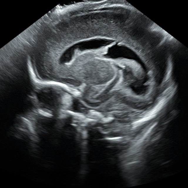
Advanced neonatal ultrasound image
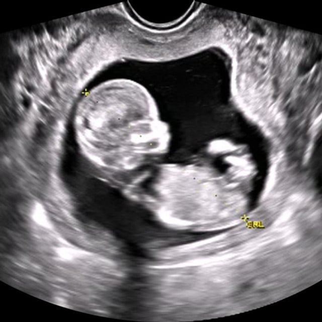
Advanced first trimester ultrasound
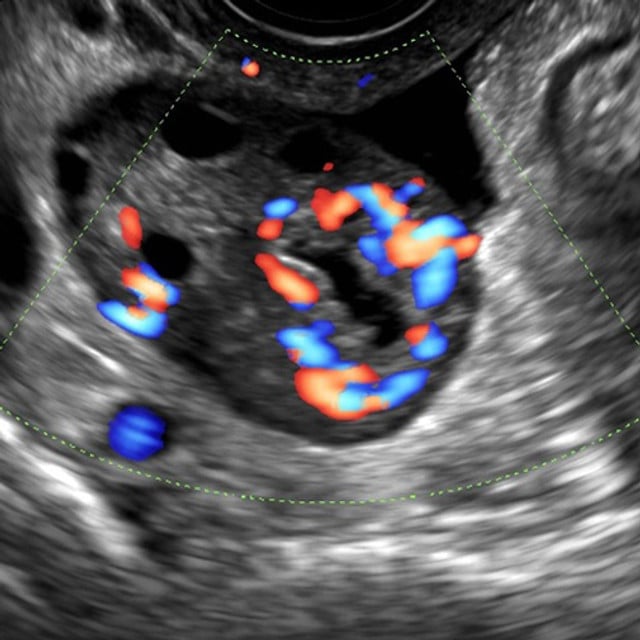
Advanced normal adnexa point of care ultrasound training scan

Advanced Clinical Series

SonoSim offers an expansive library of advanced clinical ultrasound topics to choose from. This content is geared toward clinicians looking to advance their skills in a particular clinical application.  These topics are well-suited for those who already have a strong understanding of sonography. Each SonoSim Advanced Clinical Ultrasound topic comes with an interactive, multimedia online course including ultrasound videos, knowledge checks and a Mastery Test, as well as a series of normal & pathologic scanning cases from real patients to scan in the SonoSimulator. In addition, each topic comes with CME*. This content is geared toward those looking to expand the depth and breadth of their clinical ultrasound competency.

FoCUS Advanced Clinical Ultrasound Training Part 1

Image Carrousel 24

Learn how to obtain and describe the appropriate cardiac imaging windows for the Focused Cardiac Ultrasound (FoCUS) examination to assess the heart and surrounding vascular system. Learn where, when, and how to use Doppler adjuncts to complete a FoCUS assessment. Learn to identify and evaluate unique sonographic characteristics of the heart to assess and obtain actionable measurements for cardiac output and pulmonary artery pressure.

Course Highlights

SonoSimulator Scanning Cases

The course in this module covers topics including:

  • Traditional Echocardiographic Convention
  • Ultrasound Imaging Windows for FoCUS
  • The Use of Color and Spectral Doppler in FoCUS
  • Hemodynamics
  • Pulmonary Artery Pressure Measurements
  • Case Studies including Cardiac Output and Pulmonary Hypertension
  • Tips & Pitfalls in Performing Focused Cardiac Ultrasound ( FoCUS )
  • Examination

The 10 real patient scanning cases in this module include and cover:

  • Patient Types: Adult Only, Male & Female
  • Relevant Findings/Pathology: Tricuspid Regurgitation, Aortic Regurgitation, Mitral Regurgitation and Aortic Stenosis, Dilated and Concentric Ventricular Hypertrophy, Cardiomyopathies, Pulmonary Hypertension, Use of IVC to Estimate Right Atrial Pressures
  • Doppler & Imaging Modes: Color-Flow Doppler, Continuous-Wave Doppler, Pulsed-Wave Doppler, M-Mode
  • Scanning Approaches: Parasternal, Apical, IVC, Subcostal, Suprasternal
  • Transducer Types: Phased Array
  • Special Case Features: Cardiac Calipers, Anatomic Layer Removal

How to buy5


FoCUS Advanced Clinical Ultrasound Training Part 2

Image Carrousel 25

Learn how to identify and evaluate the unique sonographic characteristics of valvular regurgitant and stenotic disorders of the heart as part of Focused Cardiac Ultrasound ( FoCUS ). Learn the ideal echocardiographic windows to use to detect valvular heart disorders. Learn to evaluate specific valvular disorders affecting the aortic, mitral, pulmonic, and tricuspid valves using ultrasound.

Course Highlights

SonoSimulator Scanning Cases

The course in this module covers topics including:

  • Anatomy & Physiology of the Heart
  • Transducer Selection & Machine Setting Review
  • Echocardiographic Technique & Ultrasound Windows
  • Doppler Imaging including Spectral and Color-Flow
  • Case Studies on Valvular Regurgitation & Valvular Stenosis, including Aortic, Mitral, Tricuspid, and Pulmonic Valve Dysfunction

The 10 real patient scanning cases in this module include and cover:

  • Patient Types: Adult Only, Male & Female
  • Relevant Findings/Pathology: Aortic Valve Regurgitation with Bicuspid Aortic Valve, Moderate Aortic Valve Regurgitation and Mild Pulmonic Valve Regurgitation, Severe Aortic Stenosis, Prosthetic Mitral Valve, Severe Mitral Valve Regurgitation and Severe Cardiomyopathy, Mitral Stenosis, Moderate-to-Severe Tricuspid Valve Regurgitation, Severe Pulmonary Hypertension with Severe Tricuspid and Pulmonary Regurgitation, Cardiac Mass, Pericardial Effusion, Pleural Effusion, and Thoracic Aortic Aneurysm, Ischemic Cardiomyopathy with Diminished LVEF
  • Doppler & Imaging Modes: Color-Flow Doppler, Continuous-Wave Doppler, Pulsed-Wave Doppler, M-Mode
  • Scanning Approaches: Parasternal, Apical, Proximal IVC, Subcostal, Suprasternal
  • Transducer Types: Phased Array
  • Special Case Features: Cardiac Calipers, Anatomic Layer Removal

How to buy5


LVAD Problem-Focused Echocardiography Advanced Clinical Ultrasound Training

Image Carrousel 45

Learn the role of bedside echocardiography in the evaluation of a Left Ventricular Assist Device ( LVAD ) patient with acute clinical decompensation. Explore the mechanics of various LVAD types, as well as LVAD complications and how to determine underlying etiology. Problem-focused echocardiographic technique is described for assessing the initial evaluation and management of LVAD patients. Develop the requisite cognitive task awareness and visuospatial skills required to perform LVAD problem-focused echocardiography.

Course Highlights

SonoSimulator Scanning Cases

The course in this module covers topics including:

  • The Role of Ultrasound in LVAD Optimization
  • LVAD Types
  • Physiologic Considerations with LVAD
  • Clinical Assessment of LVADs
  • Cardiopulmonary Resuscitation
  • LVAD-Related Complications
  • Echocardiographic Technique
  • Case Studies on Initial Resuscitation & Problem-Focused Echocardiography

The 3 real patient scanning cases in this module include and cover:

  • Patient Types: Adult Only, Male
  • Relevant Findings/Pathology: Continuous-Flow LVAD, Pericardial Effusion, Ventricular Dysfunctions, Pacemaker
  • Doppler & Imaging Modes: Color-Flow Doppler, Continuous-Wave Doppler, Power Doppler, M-Mode
  • Scanning Approaches: Parasternal, Apical, IVC Inlet, Subcostal
  • Transducer Types: Phased Array
  • Special Case Features: Additional Chest X-Ray Imaging, Anatomic Layer Removal

How to buy5


Resuscitative Transesophageal Echocardiography (TEE) Advanced Clinical Ultrasound Training

Image Carrousel 60

Learn about the use of transesophageal echocardiography (TEE) in the resuscitation of a patient in cardiac arrest. Resuscitative TEE indications, regional anatomy, echocardiographic anatomy, echocardiographic technique, and complications will be discussed. The course also reviews imaging tips, possible complications and risks, and benefits of resuscitative TEE. Develop the requisite cognitive task awareness and visuospatial skills foundation to build upon when learning how to perform TEE for resuscitation of patients in cardiac arrest.

Course Highlights

SonoSimulator Scanning Cases

The course in this module covers topics including:

  • Use of Transesophageal Echocardiography ( TEE ) in Resuscitation
  • Regional Anatomy Relevant to TEE
  • Echocardiographic Anatomy & Key Positions
  • Echocardiographic Technique & Considerations
  • Imaging Tips & Pitfalls when Performing TEE
  • TEE Case Studies of use in Acute Coronary Syndrome, Massive Pulmonary Embolism and Aortic Dissection

The 2 real patient scanning cases & 2 SkillBox cases in this module include and cover:

  • Patient Types: Adult Only, Male & Female
  • Relevant Findings/Pathology: Develop Visuospatial Skills & Cognitive Task Awareness Needed to Manipulate a TEE Transducer & Acquire the Imaging Angles Necessary to Achieve Desired TEE Views
  • Doppler & Imaging Modes: Color-Flow Doppler
  • Scanning Approaches: Intra-Oral, Upper-Esophageal, Mid-Esophageal, Transgastric, Distal-Thoracic Aorta, Mid-Thoracic Aorta, Proximal-Thoracic Aorta, Upper Esophageal Aortic Arch
  • Transducer Types: TEE
  • Special Case Features: Anatomic Layer Removal, Simulated TEE Probe Tip Flexion, Extension, Imaging Transducer Angle Changes, & Rotation Controls, Keyboard/Mouse Controls (SonoSimulator Ultrasound Probe still required for license)

How to buy5


Vascular Mapping for Preoperative Planning of Dialysis Access Advanced Clinical Ultrasound Training

Image Carrousel 80

Learn how to use ultrasound to perform vascular mapping as part of preoperative planning for dialysis access. Explore the use of ultrasound to survey upper extremity vasculature and determine the most desirable hemodialysis access site for arteriovenous fistula or graft creation. Relevant anatomy of the upper extremity vasculature, imaging protocols, and the various types of arteriovenous fistulae and grafts are discussed. Develop the requisite cognitive task awareness and psychomotor skills required for performing ultrasound vascular mapping of the upper extremities.

Course Highlights

SonoSimulator Scanning Cases

The course in this module covers topics including:

  • Vascular Mapping Indications
  • Arteriovenous Fistulae and Graft Anatomy
  • Sonographic Characteristics of Arterial and Venous Anatomy
  • Sonographic Imaging Technique Guidance for Predialysis Patients
  • Imaging Tips and Pitfalls when Vascular Mapping

The 10 real patient scanning cases in this module include and cover:

  • Patient Types: Adult Only, Male
  • Relevant Findings/Pathology: Evaluation for Calcification, Stenosis, and Thrombosis, Vascular Measurements and Mapping
  • Doppler Modes: Color-Flow, Pulsed-Wave
  • Scanning Approaches: Distal Wrist, Proximal Wrist, Antecubital Fossa, Mid-Upper Arm, Proximal Forearm, Neck
  • Transducer Types: Linear
  • Special Case Features: Compression, Anatomic Layer Removal

How to buy5


Neonatal & Infant Neurosonography Advanced Clinical Ultrasound Training

Image Carrousel 49

Learn how to perform neonatal and infant neurosonography. Get a thorough overview of the neuroanatomy of preterm and term neonates and infants, and explores the clinical indications for sonographic examination, an in-depth review of sonographic neuroanatomy and imaging techniques, and an introduction to several more commonly encountered pathologic conditions. Develop the requisite cognitive task awareness and visuospatial skills required to perform neonatal and infant neurosonography.

Course Highlights

SonoSimulator Scanning Cases

The course in this module covers topics including:

  • Anatomy & Physiology of the Neonatal & Infant Brain
  • Sonographic Neonatal & Infant Neurological Anatomy
  • Imaging Window and Planes
  • Tips & Pitfalls in Infant and Neonatal Neurosonography
  • Normal Intracranial Function of Neonates
  • Ventriculomegaly vs. Hydrocephalus

The 10 real patient scanning cases in this module include and cover:

  • Patient Types: Pediatric Only
  • Relevant Findings/Pathology: Normal Neonatal Brains, Ventriculomegaly, Hydrocephalus, Intraventricular Hemorrhage, Right Germinal Matrix Hemorrhage, Subependymal Hemorrhage, Periventricular Venous Infarction, Grade IV Right and Left Periventricular Hemorrhage, Porencephalic Cyst, Extra-Axial CSF Collections
  • Scanning Approaches: Coronal and Sagittal Anterior Fontanelle
  • Transducer Types: Phased Array
  • Special Case Features: Anatomic Layer Removal

How to buy5


Pediatric Lung Sonography Advanced Clinical Ultrasound Training

Image Carrousel 53

Learn how to use sonography to evaluate pediatric patients presenting with a variety of normal and pathologic pulmonary conditions. The sonographic characteristics of the more commonly encountered pediatric respiratory conditions will be discussed, with special emphasis on diseases of neonates and infants. Review relevant anatomy and explore the role of sonography in the evaluation of respiratory conditions such as: pneumonia, pleural effusion, pneumothorax, atelectasis, transient tachypnea, meconium aspiration syndrome, respiratory distress syndrome, bronchiolitis, and bronchopulmonary dysplasia. Develop the requisite cognitive task awareness and visuospatial skills required to perform sonographic examination of pediatric patients presenting with respiratory conditions.

Course Highlights

SonoSimulator Scanning Cases

The course in this module covers topics including:

  • Anatomy & Physiology of Pediatric Lungs
  • Sonographic Anatomy of Healthy Neonatal and Pediatric Lungs
  • Sonographic Anatomy of Pathologies such as Alveolar Interstitial Syndrome, Lung Consolidation, Atelectasis, and Pleural Effusion
  • Sonographic Techniques and Considerations in Evaluating Pediatric Lungs
  • Case Studies including Pneumonia, Respiratory Disorders, Transient Tachypnea of a Newborn, Bronchiolitis & More
  • Imaging Tips & Pitfalls when Evaluating Pediatric Lungs

The 10 real patient scanning cases in this module include and cover:

  • Patient Types: Pediatric Only
  • Relevant Findings/Pathology: Air Bronchograms, Bacterial Pneumonia, Cardiogenic Pulmonary Edema, Coalescent B-Lines (White Lung Appearance), Diffuse Air Bronchograms, Respiratory Distress Syndrome, Pleural Effusion, Pneumothorax, Severe Alveolar Interstitial Syndrome, Subpleural Consolidations, Thickened and Irregular Pleurae
  • Doppler & Imaging Modes: M-Mode
  • Scanning Approaches: Right Anterior Chest, Right Mid-Axillary Chest, Right Posterior Chest, Left Anterior Chest, Left Mid-Axillary Chest, Left Posterior Chest
  • Transducer Types: Linear, Curvilinear
  • Special Case Features: Anatomic Layer Removal

How to buy5


First-Trimester Pregnancy Advanced Clinical Ultrasound Training

Image Carrousel 23

Review traditional illustrative anatomy of the female pelvis and embryologic development alongside sonographic anatomy. Learn appropriate imaging techniques used to identify and characterize anatomic components of the female anatomy during the first-trimester of pregnancy. Learn to identify and interpret anatomy in ultrasound imaging. Identify and evaluate the unique sonographic characteristics of this phase of pregnancy, including pathology and pregnancy failure.

Course Highlights

SonoSimulator Scanning Cases

The course in this module covers topics including:

  • Anatomy of the Female Pelvis & Reproductive Tract
  • Sonographic Imaging Techniques
  • Infection Control & Safety
  • First Trimester Pregnancy Ultrasound
  • Early Gestational Development
  • Detecting Pregnancy Failure
  • Pathologic Conditions including Ectopic Pregnancy, Cesarean Scar Implantation, Subchorionic Hemorrhage, Masses & Cysts, and Molar Pregnancy
  • Twins
  • Measurements & Screenings
  • Tips & Pitfalls when Performing First-Trimester Pregnancy Ultrasound Examination

The 10 real patient scanning cases in this module include and cover:

  • Patient Types: Adult Only, Female
  • Relevant Findings/Pathology: Gestational Sacs and Yolk Sacs, Anterior and Posterior Placenta, Assessing Fetal Presentation, Bicornuate Uterus, Follicular Cysts, Corpus Luteal Cysts
  • Scanning Approaches: Transabdominal, Transvaginal
  • Transducer Types: Curvilinear, Transvaginal
  • Special Case Features: OB Calipers, Anatomic Layer Removal

How to buy5


Second and Third Trimester Pregnancy Advanced Clinical Ultrasound Training Part 1

Image Carrousel 63

Get an in-depth review of the sonographic evaluation and monitoring of normal embryologic and fetal development, including multiple pregnancies, and pathologic conditions that become evident in the second- and third-trimester of pregnancy. Develop the requisite hands-on psychomotor skills and cognitive task awareness (e.g. fetal biometry) required to optimally perform second and third trimester pregnancy ultrasound.

Course Highlights

SonoSimulator Scanning Cases

The course in this module covers topics including:

  • Sonographic Techniques for Fetal Imaging
  • Fetal Anatomy & Neuroanatomy
  • Fetal Respiratory Tract, Heart, Lungs & Diaphragm
  • Fetal Gastrointestinal, Genitourinary & Musculoskeletal Systems
  • Fetal Biometry & Pregnancy Dating
  • Cervical Length Assessment

The 10 real patient scanning cases in this module include and cover:

  • Patient Types: Adult Only, Female
  • Relevant Findings/Pathology: Determining Fetal Presentation (e.g., Cephalic vs. Breech), Fetal Biometry, Biparietal Diameter, Head Circumference, Abdominal Circumference, Femur Length, Assessing Basic Fetal Anatomy (Fetal Brain, Fetal Heart, Fetal Abdomen, Fetal Skeleton)
  • Scanning Approaches: Suprapubic, Lower Uterus, Infraumbilical, Supraumbilical
  • Transducer Types: Curvilinear
  • Special Case Features: OB Calipers, Anatomic Layer Removal

How to buy5


Second and Third Trimester Pregnancy Advanced Clinical Ultrasound Training Part 2

Image Carrousel 64

Get an in-depth review reinforcing the use of fetal biometry to evaluate fetal development of singleton and multiple pregnancies, weight discrepancies, amniotic fluid volume indexes, and maximal vertical pocket identification during the second- and third-trimesters of pregnancy. Sonographic assessment of fetal presentation, placenta, and placental location are discussed. Pathologic conditions such as intrauterine growth restriction and fetal macrosomia are described in detail. Develop the requisite hands-on psychomotor skills and cognitive task awareness required to optimally perform second and third trimester pregnancy ultrasound.

Course Highlights

SonoSimulator Scanning Cases

The course in this module covers topics including:

  • Extrafetal Anatomy
  • Fetal Lie & Presentation
  • Twin Gestation
  • Placental Evaluation & Pathology Diagnosis
  • Amniotic Fluid Assessment
  • Biophysical Profile Assessment
  • Fetal Weight
  • Intrauterine Growth Restriction
  • Fetal Macrosomia

The 10 real patient scanning cases in this module include and cover:

  • Patient Types: Adult Only, Female
  • Relevant Findings/Pathology: Determining Fetal Presentation (e.g. Cephalic vs. Breech), Fetal Biometry for Pregnancy Dating, Biparietal Diameter (Head Circumference, Abdominal Circumference, Femur Length), Assessing Basic Fetal Anatomy (Fetal Brain, Fetal Heart, Fetal Abdomen, Fetal Pelvis & Skeleton), Extrafetal Anatomy (Amniotic Fluid Index, Maximal Vertical Pocket, Placental Lie And Location)
  • Doppler & Imaging Modes: M-Mode
  • Scanning Approaches: Suprapubic, Lower Uterus, Infraumbilical, Supraumbilical, Uterine Quadrants (RUQ, RMQ, RLQ, LUQ, LMQ, LLQ)
  • Transducer Types: Curvilinear
  • Special Case Features: OB Calipers, Anatomic Layer Removal

How to buy5


GYN Ultrasound of Nonpregnant Normal Uterus Advanced Clinical Training

Image Carrousel 34

Learn to identify and evaluate a nonpregnant female uterus and pelvis using ultrasonography, including anatomy, sonographic characteristics and ultrasound imaging techniques. Learn to recognize and evaluate normal variations of uterine appearance and positioning in the nonpregnant female, with sonography.

Course Highlights

SonoSimulator Scanning Cases

The course in this module covers topics including:

  • Indications for Ultrasound Examination
  • Normal Gynecologic Anatomy
  • Female Pelvis Physiology
  • Transabdominal, Transvaginal Sonographic Techniques
  • Uterine Positions
  • Uterus Evaluation & Measurement
  • Case Studies of Posterior Cul-de-Sac Free Fluid, and Intrauterine Device ( IUD )

The 10 real patient scanning cases in this module include and cover:

  • Patient Types: Adult Only, Female
  • Relevant Findings/Pathology: Varying Uterine Fundal Positioning (Anteverted, Anteflexed, Retroverted, Retroflexed), Nabothian Cysts, Physiology vs. Pathologic Fluid Screening, IUD-Placement Assessment, Assessment of Endometrial Phasic Changes, Color Doppler Application on Adnexal Region
  • Doppler & Imaging Modes: Color-Flow Doppler
  • Scanning Approaches: Transabdominal, Transvaginal
  • Transducer Types: Curvilinear, Transvaginal
  • Special Case Features: Anatomic Layer Removal

How to buy5


GYN Ultrasound of Nonpregnant Abnormal Uterus Advanced Clinical Training Part 1

Image Carrousel 32

Learn to identify and evaluate the nonpregnant uterus, including anatomy, sonographic characteristics and imaging techniques. Understand how to evaluate the abnormal, nonpregnant uterus with ultrasonography. Learn to recognize and evaluate various abnormal conditions of the myometrium in the nonpregnant female, using ultrasound.

Course Highlights

SonoSimulator Scanning Cases

The course in this module covers topics including:

  • Introduction to Abnormal Uterus Ultrasound
  • Sonographic Image Acquisition of the Uterus
  • Congenital Uterine Malformations
  • Sonographic Evaluation of Adenomyosis
  • Sonographic Evaluation of Leiomyomas
  • Localization of Intrauterine Devices ( IUDs )

The 10 real patient scanning cases in this module include and cover:

  • Patient Types: Adult Only, Female
  • Relevant Findings/Pathology: Congenital Uterine Malformations, IUD-Placement Assessment, Adenomyosis and Leiomyomas, Color Doppler Application on Adnexal Region
  • Doppler & Imaging Modes: Color-Flow Doppler
  • Scanning Approaches: Transabdominal, Transvaginal
  • Transducer Types: Curvilinear, Transvaginal
  • Special Case Features: Anatomic Layer Removal

How to buy5


GYN Ultrasound of Nonpregnant Abnormal Uterus Advanced Clinical Training Part 2

Image Carrousel 33

Learn to identify and evaluate various endometrial conditions in the nonpregnant abnormal uterus, including sonographic characteristics, and imaging techniques.

Course Highlights

SonoSimulator Scanning Cases

The course in this module covers topics including:

  • Introduction to Abnormal Uterine Bleeding
  • Sonographic Imaging Techniques for Evaluating the Endometrium
  • Case Studies including Endometrial Polyps, Submucosal Leiomyomas, Endometrial Adhesions, Subinvolution of the Placental Site, Endometrial Cancer, and Tamoxifen Effect on Endometrium

The 5 real patient scanning cases in this module include and cover:

  • Patient Types: Adult Only, Female
  • Relevant Findings/Pathology: Endometrial Adhesions, Retained Products of Conception, Endometrial Polyps, Leiomyomas, Pathologic Free Fluid In and Around Uterus, Color Doppler Evaluation of Adnexal Structures
  • Doppler & Imaging Modes: Color-Flow Doppler
  • Scanning Approaches: Transabdominal, Transvaginal
  • Transducer Types: Curvilinear, Transvaginal
  • Special Case Features: Anatomic Layer Removal

How to buy5


GYN Ultrasound Normal Adnexa Advanced Clinical Training

Image Carrousel 31

Learn to identify and evaluate the normal adnexa, including anatomy, unique sonographic characteristics and ultrasound imaging techniques used in evaluating the normal adnexa. Learn to identify and evaluate normal variations of appearance and positioning of the normal adnexa.

Course Highlights

SonoSimulator Scanning Cases

The course in this module covers topics including:

  • Gynecologic Ultrasound of Normal Adnexa
  • Adnexal Anatomy
  • Female Pelvis Physiology, including Menstruation and Ovarian Cycles
  • Sonographic Anatomy of the Adnexa
  • Ultrasound Imaging Techniques for Ovary and Fallopian Tube Evaluation
  • Adnexal Masses Case Studies including Hemorrhagic Cyst

The 10 real patient scanning cases in this module include and cover:

  • Patient Types: Adult Only, Female
  • Relevant Findings/Pathology: Postpubertal Patient Cases, Postmenopausal Patient Cases, Menstrual Cycle-Related Ovarian Changes (Ovarian Follicles, Ovarian Follicular Cysts, Corpus Luteum, Posterior Cul-De-Sac Physiologic Free Fluid)
  • Doppler & Imaging Modes: Color-Flow Doppler
  • Scanning Approaches: Transabdominal, Transvaginal
  • Transducer Types: Curvilinear, Transvaginal
  • Special Case Features: Anatomic Layer Removal

How to buy5


GYN Ultrasound Nonmalignant Adnexal Conditions Advanced Clinical Training

Image Carrousel 30

Learn to evaluate adnexal masses using ultrasound, including imaging techniques used in sonographic evaluation of nonmalignant adnexal conditions. Understand the implications of ultrasound findings for specific gynecological conditions. Learn to identify and evaluate nonmalignant adnexal conditions, such as hydrosalpinx, tubo-ovarian abscess, and adnexal torsion, using ultrasonography.

Course Highlights

SonoSimulator Scanning Cases

The course in this module covers topics including:

  • Introduction to Ultrasound of Nonmalignant Adnexal Conditions
  • Adnexal Anatomy Overview
  • Various Types of Ovarian Cysts, including Dermoid, Ovarian Fibroma, Hemorrhagic, and Polycystic Ovary Syndrome
  • Case Studies of Pedunculated Leiomyoma, Hydrosalpinx, Tubo-Ovarian Abscess, Peritoneal Inclusion Cyst, Paraovarian Cyst and Adnexal Torsion

The 10 real patient scanning cases in this module include and cover:

  • Patient Types: Adult Only, Female
  • Relevant Findings/Pathology: Simple & Complex Ovarian Cysts, Hemorrhagic Cysts, Endometriomas, Dermoid Cysts, Hydrosalpinx
  • Doppler & Imaging Modes: Color-Flow Doppler
  • Scanning Approaches: Transabdominal, Transvaginal
  • Transducer Types: Curvilinear, Transvaginal
  • Special Case Features: Anatomic Layer Removal

How to buy5


GYN Ultrasound Malignant Adnexal Conditions Advanced Clinical Training

Image Carrousel 29

Learn to evaluate adnexal masses using ultrasound, including imaging techniques used in sonographic evaluation of malignant adnexal conditions. Learn to identify the risk factors for ovarian cancer and evaluate ovarian cancer via ultrasonography.

Course Highlights

SonoSimulator Scanning Cases

The course in this module covers topics including:

  • Introduction to Malignant Adnexal Conditions
  • Adnexal Anatomy
  • Evaluation of Adnexal Masses for Malignancy
  • Sonographic Techniques including Doppler Flow
  • Sonographic Characterization of Adnexal Masses
  • Borderline Tumors
  • Ovarian Cancer Overview
  • Ovarian Cancer Screening

The 10 real patient scanning cases in this module include and cover:

  • Patient Types: Adult Only, Female
  • Relevant Findings/Pathology: Complex Adnexal Cysts, Germ Cell Tumors, Leiomyomas, Pathologic Free Fluid, Dermoid Cysts, Endometriomas, Ovarian Fibromas
  • Doppler & Imaging Modes: Color-Flow Doppler
  • Scanning Approaches: Transabdominal, Transvaginal
  • Transducer Types: Curvilinear, Transvaginal
  • Special Case Features: Anatomic Layer Removal

How to buy5

 
Peripheral ultrasound image for point of care ultrasound training applications
Procedure femoral ultrasound scan
Procedural IJ ultrasound
Paracentesis procedure ultrasound training scan
Glenohumeral procedure ultrasound scan
Tibiotalar ultrasound-guided procedure in SonoSim's point of care ultrasound training

Ultrasound-Guided Procedure Series

SonoSim offers an extensive selection of ultrasound-guided procedure training options. This content is geared toward clinicians looking to advance their skills in specific needle-based procedures. Each SonoSim Ultrasound-Guided Procedure topic comes with an interactive, multimedia online course including ultrasound videos, knowledge checks and a Mastery Test, as well as scanning cases of real patient anatomy to practice on in the SonoSimulator. The SonoSimulator helps develop image acquisition & interpretation, and needle visualization for both in- and out-of-plane guidance, and real-time guidance with feedback.

SonoSim's proprietary platform seamlessly combines real patient anatomy with interactive simulation, allowing you to immerse yourself in the world of ultrasound anatomy and uncover the intricacies of imaging artifacts. Discover the art of applying Doppler techniques when appropriate, as you navigate through our comprehensive ultrasound-guided procedure training. A standout feature of SonoSim's training is the integration of expert-trained artificial intelligence (AI) that provides instant feedback on needle trajectory and procedural success. Enhancing your learning experience further, our informative video tutorials serve as an on-demand virtual coach, acting as a personal guide every step of the way.

In addition, each topic comes with CME*. This content is geared toward those looking to increase safety and effectiveness of common needle-based procedures that benefit from ultrasound guidance.

Ultrasound-Guided Procedures Introductory Training

Image Carrousel 77

Learn the basic principles of performing ultrasound-guided procedures. Develop the requisite hands-on psychomotor skills and cognitive task awareness required to optimally perform ultrasound-guided procedures. Included SkillBox scanning cases provide risk-free cognitive task training opportunities.

Course Highlights

SonoSimulator Scanning Cases

The course in this module covers topics including:

  • Procedure Indications and Contraindications
  • Site Selection
  • Sonography Anatomy & Characteristics
  • Transducer Selection & Settings
  • Ergonomic Considerations
  • Patient Positioning
  • Infection Prevention
  • Survey Scan & Procedure Steps
  • Doppler-Mode Imaging
  • Tips, Pitfalls, & Complications

The 10 SkillBox scanning cases in this module include:

  • 20 Knowledge Check Questions
  • Real Time Assessment & Feedback
  • A 20 Question Mastery Test

How to buy5


Peripheral Venous Access Ultrasound-Guided Procedure Training

Image Carrousel 54

Learn how to use sonography to guide peripheral venous access in patients. The needle-based peripheral venous access procedure feature provides cognitive task training on ultrasound-guided needle-based procedures. Ultrasound-guided peripheral venous access procedure indications, sonographic techniques and scanning approaches, including imaging tips, pitfalls, and procedure complications are described.

Course Highlights

SonoSimulator Scanning Cases

The course in this module covers topics including:

  • Procedure Indications & Contraindications
  • Site Selection
  • Sonography Anatomy & Characteristics
  • Transducer Selection & Settings
  • Ergonomic Considerations
  • Patient Positioning
  • Ergonomic Considerations
  • Infection Prevention
  • Survey Scan & Procedure Steps
  • Doppler-Mode Imaging
  • Tips, Pitfalls & Complications

The 10 real patient scanning cases in this module include and cover:

  • Patient Types: Male & Female, 7 Adult & 3 Pediatric
  • Relevant Findings/Pathology: Variable Body Morphology, Right and Left Arms, Dorsal Metacarpal Veins, Cephalic Veins, Brachial Veins, Basilic Veins, Median Cubital Vein
  • Scanning Approaches: Short Axis & Long Axis
  • Doppler & Imaging Modes: Power Doppler
  • Procedure Techniques: In-Plane & Out-of-Plane Needle Guidance
  • Transducer Types: Linear
  • Special Case Features: Compression

How to buy5


Femoral Line Placement Ultrasound-Guided Procedure Training

Image Carrousel 22

Get an in-depth understanding of the basic principles of how to perform ultrasound-guided femoral central line placement. Develop the requisite hands-on psychomotor skills and cognitive task awareness required to optimally perform femoral vein access with the aid of ultrasound.

Course Highlights

SonoSimulator Scanning Cases

The course in this module covers topics including:

  • Procedure Indications and Contraindications
  • Site Selection
  • Sonographic Anatomy & Characteristics
  • Transducer Selection & Settings
  • Ergonomic Considerations
  • Patient Positioning
  • Infection Prevention
  • Survey Scan and Procedure Steps
  • Doppler-Mode Imaging
  • Tips, Pitfalls, and Complications

The 10 real patient scanning cases in this module include and cover:

  • Patient Types: Adult Only, Male & Female
  • Relevant Findings/Pathology: Normal Patient Anatomy, Variable Body Habitus, Variable Hemodynamic and Hydration Status
  • Doppler & Imaging Modes: Power Doppler
  • Special Case Features: Compressions

How to buy5


Internal Jugular Vein Cannulation Ultrasound-Guided Procedure Training

Image Carrousel 38

Get an in-depth understanding of the basic principles of how to perform ultrasound-guided internal jugular vein cannulation. Develop the requisite hands-on psychomotor skills and cognitive task awareness required to optimally perform ultrasound-guided internal jugular vein cannulation. Ultrasound-guided internal jugular vein cannulation procedure indications, sonographic techniques and scanning approaches, including imaging tips, pitfalls, and procedure complications are also provided.

Course Highlights

SonoSimulator Scanning Cases

The course in this module covers topics including:

  • Procedure Indications & Contraindications
  • Site Selection
  • Sonography Anatomy & Characteristics
  • Transducer Selection & Settings
  • Ergonomic Considerations
  • Patient Positioning
  • Infection Prevention
  • Survey Scan & Procedure Steps
  • Doppler-Mode Imaging
  • Tips, Pitfalls, & Complications

The 10 real patient scanning cases in this module include and cover:

  • Patient Types: Adult Only, Male & Female
  • Relevant Findings/Pathology: Variable Body Morphologies, Right and Left Neck, Variable Hemodynamic & Hydration Status
  • Doppler & Imaging Modes: Power Doppler
  • Scanning Approaches: Transverse & Longitudinal
  • Procedural Techniques: In-Plane & Out-of-Plane Needle Guidance
  • Transducer Types: Linear
  • Special Case Features: Compression, Anatomic Layer Removal

How to buy5


Subclavian Vein Cannulation Ultrasound-Guided Procedure Training

Image Carrousel 71y

Get in-depth understanding of the basic principles of how to perform ultrasound-guided subclavian vein cannulation. Develop the requisite hands-on psychomotor skills and cognitive task awareness required to optimally perform ultrasound-guided subclavian vein cannulation. Ultrasound-guided subclavian vein cannulation procedure indications, sonographic techniques and scanning approaches, including imaging tips, pitfalls, and procedure complications are also provided.

Course Highlights

SonoSimulator Scanning Cases

The course in this module covers topics including:

  • Procedure Indications & Contraindications
  • Site Selection
  • Sonography Anatomy & Characteristics
  • Transducer Selection & Settings
  • Ergonomic Considerations
  • Patient Positioning
  • Infection Prevention
  • Survey Scan & Procedure Steps
  • Doppler-Mode Imaging
  • Tips, Pitfalls, & Complications

The 10 real patient scanning cases in this module include and cover:

  • Patient Types: Adult Only, Male & Female
  • Relevant Findings/Pathology: Variable Body Morphologies, Right and Left Neck, Variable Hemodynamic & Hydration Status
  • Doppler & Imaging Modes: Power Doppler, Color-Flow Doppler, Pulsed-Wave Doppler
  • Scanning Approaches: Infraclavicular
  • Procedure Techniques: In-Plane & Out-of-Plane Needle Guidance
  • Transducer Types: Linear

How to buy5


Ultrasound-Guided Paracentesis Procedure Training

Image Carrousel 75

Learn how to perform an ultrasound-guided paracentesis. This course explores the use of ultrasound to detect ascites, regional anatomy, clinical indications for performing paracentesis, procedural complications, and procedural steps for performing ultrasound-guided paracentesis. Develop the requisite cognitive task awareness and visuospatial skills required to perform ultrasound-guided paracentesis.

Course Highlights

SonoSimulator Scanning Cases

The course in this module covers topics including:

  • Procedure Indications & Contraindications
  • Site Selection
  • Sonography Anatomy & Characteristics
  • Transducer Selection & Settings
  • Ergonomic Considerations
  • Patient Positioning
  • Infection Prevention
  • Survey Scan & Procedure Steps
  • Doppler-Mode Imaging
  • Tips, Pitfalls, & Complications

The 10 real patient cases in this module include and cover:

  • Patient Types: Adult Only, Male & Female
  • Relevant Findings/Pathology: Variable Body Morphologies, Small-Volume Ascites Cases, Moderate-Volume Ascites Cases, Large-Volume Ascites Cases, Male and Female Patients with Variable Body Habitus, Multiple Diagnostic Imaging Windows, Color-Flow Doppler Screening of Overlying Abdominal Wall
  • Doppler & Imaging Modes: Color Doppler
  • Procedural Techniques: In-Plane & Out-of-Plane Needle Guidance
  • Transducer Types: Curvilinear and Linear-Array Transducers

How to buy5


Ultrasound-Guided Pericardiocentesis Procedure Training

Image Carrousel 76

Learn the principles of performing an ultrasound-guided pericardiocentesis. Explore the use of ultrasound to detect pericardial effusions, understand clinical indications for performing pericardiocentesis, and how to identify echocardiographic findings suggestive of cardiac tamponade physiology. Learn the steps necessary for performing ultrasound-guided pericardiocentesis. Develop the requisite cognitive task awareness and visuospatial skills required to perform ultrasound-guided pericardiocentesis.

Course Highlights

SonoSimulator Scanning Cases

The course in this module covers topics including:

  • Procedure Indications & Contraindications
  • Site Selection
  • Sonography Anatomy & Characteristics
  • Transducer Selection & Settings
  • Ergonomic Considerations
  • Patient Positioning
  • Infection Prevention
  • Survey Scan & Procedure Steps
  • Doppler-Mode Imaging
  • Tips, Pitfalls, & Complications

The 10 real patient scanning cases in this module include and cover:

  • Patient Types: Male & Female, 10 Adult
  • Relevant Findings/Pathology: Small Pericardial Effusions, Moderate Pericardial Effusions, Large Pericardial Effusions, Cardiac Tamponade Cases, Male & Female Patients with Variable Body Habitus, Multiple Diagnostic Imaging Windows
  • Doppler & Imaging Modes: M-Mode
  • Scanning Approaches: Parasternal, Apical, & Subcostal Windows
  • Procedure Techniques: In-Plane & Out-of-Plane Needle Guidance

How to buy5


Ultrasound-Guided Thoracentesis Procedure Training

Image Carrousel 78

Learn how to perform an ultrasound-guided thoracentesis. The course explores the use of ultrasound to detect pleural effusions, clinical indications for performing a thoracentesis, procedural complications, and procedural steps for performing ultrasound-guided thoracentesis. Develop the requisite cognitive task awareness and visuospatial skills required to perform ultrasound-guided thoracentesis.

Course Highlights

SonoSimulator Scanning Cases

The course in this module covers topics including:

  • Procedure Indications & Contraindications
  • Site Selection
  • Sonography Anatomy & Characteristics
  • Transducer Selection & Settings
  • Ergonomic Considerations
  • Patient Positioning
  • Infection Prevention
  • Survey Scan & Procedure Steps
  • Doppler-Mode Imaging
  • Tips, Pitfalls, & Complications

The 10 real patient scanning cases in this module include and cover:

  • Patient Types: Adult Only, Male & Female
  • Relevant Findings/Pathology: Small-volume Pleural Effusion Cases, Moderate-Volume Pleural Effusion Cases, Large-Volume Pleural Effusion Cases, Underlying lung pathology (Alveolar-Interstitial Syndrome, Lung Adhesions, Pulmonary Fibrosis), Male and Female Patients with Variable Body Habitus
  • Doppler & Imaging Modes: M-Mode and Color-Flow Doppler Screening of Overlying Soft Tissue
  • Scanning Approaches: Posterolateral
  • Procedural Techniques: In-Plane & Out-of-Plane Needle Guidance
  • Transducer Types: Curvilinear, Linear-Array

How to buy5


Biceps Tendon Sheath Injection Ultrasound-Guided Procedure Training

Image Carrousel 10

Learn both the regional and sonographic anatomy and how to interpret ultrasound imagery of the shoulder joint. Learn sonographic techniques and scanning approaches used in ultrasound-guided biceps tendon sheath injection. Learn to evaluate various biceps tendon sheath pathologies with ultrasonography and clinical indications for performing biceps tendon sheath injections.

Course Highlights

SonoSimulator Scanning Cases

The course in this module covers topics including:

  • Procedure Indications & Contraindications
  • Site Selection
  • Sonographic Anatomy & Characteristics
  • Transducer Selection & Settings
  • Ergonomic Considerations
  • Patient Positioning
  • Infection Prevention
  • Survey Scan & Procedure Steps
  • Tips, Pitfalls & Complications

The real adult patient training case in this module includes and covers:

  • Patient Type: Adult Female
  • Doppler & Imaging Modes: Greyscale & Color Doppler imaging
  • Scanning Approaches: Lateral-to-Medial and Proximal-to-Distal
  • Procedural Techniques: In-Plane, Out-of-Plane, & combination
  • Transducer Types: Linear

How to buy5


Plantar Fasciitis Ultrasound-Guided Injection Procedures

Image Carrousel 55

Learn the basic principles of performing ultrasound-guided plantar intrafascial & perifascial injections. Review relevant anatomic landmarks and the use of ultrasound to evaluate the plantar fascia. Learn commonly agreed upon indications for performing this procedure. Develop cognitive task awareness and visuospatial skills required to perform ultrasound-guided injections to alleviate plantar fasciitis.

Course Highlights

SonoSimulator Scanning Cases

The course in this module covers topics including:

  • Plantar Fascial Anatomy & Physiology
  • Plantar Fascial Disorders
  • Vascular Supply & Nerves
  • Sonography Anatomy & Characteristics
  • Pathologic Conditions
  • Procedure Indications & Contraindications
  • Transducer Selection & Patient Positioning
  • Survey Scan & Procedure Steps
  • Alternative Approaches
  • Tips, Pitfalls, & Complications

The 2 real patient scanning cases in this module include and cover:

  • Patient Type: 1 Adult Male; 1 Adult Female
  • Relevant Findings/Pathology: Normal Proximal Plantar Fascia; Pathologic Thickened Proximal Plantar Fascia
  • Doppler & Imaging Modes: Color Doppler
  • Transducer Types: Linear
  • Scanning Approaches: Plantar Intrafascial Injection - Medial-to-Lateral Procedural Approach; Plantar Perifascial Injection - Medial-to-Lateral Procedural Approach
  • Procedural Techniques: In-Plane

How to buy5


Acromioclavicular Joint Injection & Aspiration Ultrasound-Guided Procedure Training

Image Carrousel 1

Learn both the regional and sonographic anatomy of the acromioclavicular joint and how to interpret ultrasound imagery of this joint. Learn sonographic techniques and scanning approaches used in ultrasound-guided acromioclavicular joint injection and aspiration. Learn to evaluate various acromioclavicular joint pathologies with ultrasonography and clinical indications for performing acromioclavicular joint injections and acromioclavicular joint aspiration.

Course Highlights

SonoSimulator Scanning Cases

The course in this module covers topics including:

  • Procedure Indications & Contraindications
  • Site Selection
  • Sonography Anatomy & Characteristics
  • Transducer Selection & Settings
  • Ergonomic Considerations
  • Patient Positioning
  • Infection Prevention
  • Survey Scan & Procedure Steps
  • Doppler-Mode Imaging
  • Tips, Pitfalls, & Complications

The one (1) real patient scanning case in this module includes and covers:

  • Patient Type: Adult Female
  • Relevant Findings/Pathology: One Real Adult Patient Training Case
  • Transducer Types: Linear-array
  • Scanning Approaches: Lateral-to-Medial and Anterior-to-Posterior
  • Procedural Techniques: In-Plane, Out-of-Plane, and Combination Techniques

How to buy5


Glenohumeral Joint Injection & Aspiration Ultrasound-Guided Procedure Training

Image Carrousel 28

Learn the relationship between traditional illustrative anatomy and sonographic anatomy of the glenohumeral joint. Learn how glenohumeral joint injections and glenohumeral joint aspiration is performed, including the location and appearance of critical anatomical components in ultrasound imaging. Sonographic techniques and scanning approaches used in performing ultrasound-guided glenohumeral joint injection and aspiration procedures are covered. Ultrasound-guided glenohumeral joint injection & aspiration procedure indications, sonographic techniques and scanning approaches, including imaging tips, pitfalls, and procedure complications are covered.

Course Highlights

SonoSimulator Scanning Cases

The course in this module covers topics including:

  • Procedure Indications & Contraindications
  • Site Selection
  • Sonographic Anatomy
  • Sonography Anatomy & Characteristics
  • Transducer Selection & Settings
  • Ergonomic Considerations
  • Patient Positioning
  • Infection Prevention
  • Survey Scan & Procedure Steps
  • Tips, Pitfalls & Complications

The real patient training case in this module includes and covers:

  • Patient Types: Adult Female
  • Scanning Approaches: Anterior & Posterior Glenohumeral Joint
  • Procedural Techniques: In-Plane Needle Guidance
  • Transducer Types: Linear Array

How to buy5


Subacromial-Subdeltoid Bursa Injection & Aspiration Ultrasound-Guided Procedure Training

Image Carrousel 70

Get an in-depth understanding of the basic principles of how to perform ultrasound-guided subacromial-subdeltoid bursal injection and aspiration. The course explores the use of ultrasound to detect bursal fluid and clinical indications for performing the procedure. Subacromial-subdeltoid bursitis, subacromial impingement syndrome, and rotator cuff injuries are discussed. Develop the requisite cognitive task awareness and visuospatial skills required to perform ultrasound-guided subacromial-subdeltoid bursa injection and aspiration. Ultrasound-guided subacromial-subdeltoid bursa injection and aspiration procedure indications, sonographic techniques and scanning approaches, including imaging tips, pitfalls, and procedure complications are described.

Course Highlights

SonoSimulator Scanning Cases

The course in this module covers topics including:

  • Procedure Indications & Contraindications
  • Site Selection
  • Sonography Anatomy & Characteristics
  • Transducer Selection & Settings
  • Ergonomic Considerations
  • Patient Positioning
  • Infection Prevention
  • Survey Scan & Procedure Steps
  • Doppler-Mode Imaging
  • Tips, Pitfalls & Complications

The real patient scanning case in this module includes and covers:

  • Patient Types: Adult Female
  • Relevant Findings/Pathology: One Subacromial-Subdeltoid Bursa Case
  • Scanning Approaches: Lateral-to-Medial Approach
  • Procedural Techniques: In-Plane Needle Guidance

How to buy5


Tibiotalar Joint Injection & Aspiration Ultrasound-Guided Procedure Training

Image Carrousel 73

Get an in-depth understanding of the basic principles of how to perform ultrasound-guided tibiotalar joint injection and/or aspiration. Review relevant anatomy and explore the use of ultrasound to evaluate tibiotalar joint pathologies and clinical indications for performing the procedure. Acute and chronic pathologic conditions that afflict the tibiotalar joint are discussed, including trauma, degenerative arthritis, cartilaginous lesions, inflammatory conditions, and infections. Develop the requisite cognitive task awareness and visuospatial skills required to perform ultrasound-guided tibiotalar joint injection.

Course Highlights

SonoSimulator Scanning Cases

The course in this module covers topics including:

  • Procedure Indications & Contraindications
  • Site Selection
  • Sonography Anatomy & Characteristics
  • Transducer Selection & Settings
  • Ergonomic Considerations
  • Patient Positioning
  • Infection Prevention
  • Survey Scan & Procedure Steps
  • Doppler-Mode Imaging
  • Tips, Pitfalls & Complications

The real patient training case in this module includes and covers:

  • Patient Types: Adult Male
  • Relevant Findings/Pathology: One Training Case Featuring Real Patient Anatomy
  • Doppler & Imaging Modes: Color Doppler
  • Scanning Approaches: Medial-to-Lateral Procedural Approach
  • Procedural Techniques: In-Plane Needle Guidance
  • Transducer Types: Linear

How to buy5


Metatarsophalangeal Joint Injection & Aspiration Ultrasound-Guided Procedure Training

Image Carrousel 46

Learn the basic principles of performing ultrasound-guided metatarsophalangeal joint injection and aspiration. Review relevant anatomy and the use of ultrasound to evaluate metatarsophalangeal joint pathologies. Learn the clinical indications for performing the procedure. Review acute and chronic pathologic conditions that afflict the metatarsophalangeal joint, including trauma, degenerative arthritis, inflammatory conditions, infections, and masses. Develop cognitive task awareness and visuospatial skills required to perform ultrasound-guided metatarsophalangeal joint injection.

Course Highlights

SonoSimulator Scanning Cases

The course in this module covers topics including:

  • Metatarsophalangeal Joint Disorders
  • Metatarsophalangeal Anatomy & Physiology
  • Arterial Supply & Nerves
  • Sonography Anatomy & Characteristics
  • Pathologic Conditions, Including Synovitis & Joint Effusion
  • Procedure Indications & Contraindications
  • Transducer Selection & Patient Positioning
  • Survey Scan & Procedure Steps
  • Alternative Approaches
  • Tips, Pitfalls, & Complications

The 2 real patient scanning cases in this module include and cover:

  • Patient Types: 2 Adult Males
  • Relevant Findings/Pathology: Right Foot Normal; Right Foot Severe Osteoarthritis
  • Transducer Types: Linear
  • Scanning Approaches: Dorsal, Medial, and Plantar; Proximal-to-Distal
  • Procedural Techniques: In-Plane

How to buy5


Neonatal & Infant Lumbar Puncture Ultrasound-Guided Procedure Training

Image Carrousel 49-1

Gain an in-depth understanding of the basic principles of performing ultrasound-guided neonatal and infant lumbar punctures. Review relevant anatomic landmarks that are instrumental to performing ultrasound-guided lumbar punctures. Develop cognitive task awareness and visuospatial skills required to perform the procedure to evaluate for sepsis or central nervous system infections. Review the anatomy of the neonatal and infant spine, and commonly agreed-upon clinical indications for performing ultrasound-guided lumbar punctures. Learn optimal transducer selection, patient positioning, imaging approaches, and sonographic techniques for procedure execution. Imaging tips and pitfalls, and potential procedure complications are discussed.

Course Highlights

SonoSimulator Scanning Cases

The course in this module covers topics including:

  • Neonate/Infant Lumbar Puncture Procedure Indications
  • Infant & Neonate Spinal Anatomy
  • Sonographic Pediatric Anatomy & Characteristics
  • Procedure Preparation
  • Transducer Selection & Patient Positioning
  • Survey Scan & Procedure Steps
  • Tips, Pitfalls, & Complications
 

The 2 real patient scanning cases in this module include and cover:

  • Patient Type: 2 Infant
  • Relevant Findings/Pathology: Conus terminating at L2 and L3
  • Doppler & Imaging Modes: Color Doppler
  • Transducer Types: Linear
  • Scanning Approaches: Dorsal
  • Procedural Techniques: In-Plane & Out-of-Plane Needle Guidance

How to buy5


Explore the SonoSim Ecosystem

Study
Tools
Study ultrasound training

Case-based learning & reference tools advance training on-the-go

Apply
SonoSim LiveScan
Apply ultrasound training in medical decision-making scenarios with LiveScan

Apply U/S knowledge in medical decision-making

Integrate
Resources
Integration of SonoSim into a point of care ultrasound training program

Integrate ultrasound into curricula

Track
Performance Tracker
Instructor tracking learner progress in point of care ultrasound training program

Streamline performance tracking & image review

Click me
Practice
Procedures
Practice ultrasound procedures for point of care ultrasound applications

U/S-guided procedure training with real-time feedback

Learn
Courses
Learn with courses and enrich your ultrasound education

On-demand access to world-class ultrasound educators

Bedside Clinical Practice

SonoSim complements bedside learning & accelerates the path to ultrasound competency.

SCHEDULE A DEMO
Scan
SonoSimulator
Learner scanning in the SonoSimulator for ultrasound training

Provide unlimited scanning of real patient pathology

How to Buy

Best Value_blue_v3

Schools &
Educational Groups

academic

Interactive, Hands-On, Virtual Learning • Textbook Pricing

Unlimited Access Licenses 
DMS/UME/GME Discount
less than $50/mo/learner
 

  • All Ultrasound Topics
  • SonoSimulator Software
  • All Normal & Pathologic Cases
  • Tracking & Image Portfolios
  • QuestionBank & SPI Test Prep
  • Integration Support
SONOSIM FOR PROGRAMS

++ CME* add-on available
++SonoSim LiveScan add-on available

List price per non-transferrable seat license, based on 3-year term, 5 learner minimum. Alternate contract terms available on request. Billed annually. Licenses transferrable between learners each year.  Additional start-up costs include onboarding and a SonoSimulator probe hardware purchase of $249 each.

Hospitals & 
Medical Groups

hospitals

Self-Paced, Remote Ultrasound Learning • Save on CME & Travel

Unlimited Access for Practicing Physicians, Nurses, PAs, NPs, EMTs...
as low as $799/learner

  • All Ultrasound Topics
  • SonoSimulator Software
  • All Normal & Pathologic Cases
  • Tracking & Image Portfolios
  • QuestionBank & SPI Test Prep
  • Integration Support
SONOSIM FOR GROUPS

++ CME* available add-on available
++ SonoSim LiveScan add-on available

List price per non-transferrable seat license, based on 3-year term, 5 learner minimum. Alternate contract terms available on request. Billed annually. Licenses transferrable between learners each year.  Additional start-up costs include onboarding and a SonoSimulator probe hardware purchase of $249 each.

Students &
Recent Graduates

student

Lead Your Peers
Self-Paced Ultrasound Learning

Annual Memberships
for Aspiring Providers
$599/year

  • All Ultrasound Topics
  • SonoSimulator Software
  • All Normal & Pathologic Cases
  • Certificates & Image Portfolios
  • QuestionBank & SPI Test Prep
SONOSIM FOR STUDENTS

Not eligible for CME. Proof of academic status required. Renewable annually as long as eligibility applies. Upgradeable to Provider license 1+ year post-graduation. 

Individual Medical Providers

healthcare

Build the Ultrasound Skills You Need • Get CME

Flexible Packages • CME included
Choose from 80+ Topics
Each Topic from $399 & up

  • Choose Your Ultrasound Topics
  • SonoSimulator Software
  • Relevant Scanning Cases
  • Image Portfolio
  • Up to 6 CME credits per topic*
SONOSIM FOR PROVIDERS

CME included. All your topics renewable annually for $249. Exclusive member discounts & benefits apply throughout the year. 

SonoSim ultrasound training ecosystem elements

Let's Talk About Ultrasound Training

Contact Us

The SonoSim Wave
An Ultrasound Insights Newsletter

Get the latest trends in ultrasound training, education, & applications delivered to your inbox.
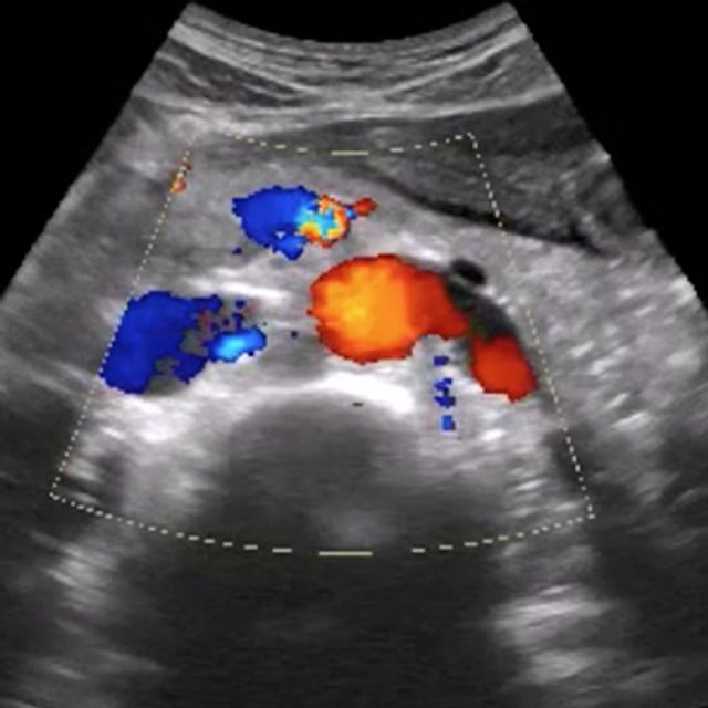
1 Tolsgaard MG, Todsen T, Sorensen JL, et al. International multispecialty consensus on how to evaluate ultrasound competence: a Delphi consensus survey. PLoS One. 2013;8(2):e57687. doi: 10.1371/journal.pone.0057687. Epub 2013 Feb 28. PMID: 23469051; PMCID: PMC3585207.