If you’re curious about the types of ultrasound probes and their uses, we've compiled them for you here. SonoSim is dedicated to helping students, academic & DMS programs, and medical providers overcome the barriers to ultrasound training. We hope to hear from you if you’re interested in The Easiest Way to Learn & Teach UltrasoundⓇ!
What are Ultrasound Probes?
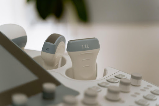
An ultrasound probe, also called an ultrasound transducer, is a device that produces sound waves that bounce off body tissue to make echoes. The transducer receives the echoes and sends them to a computer that uses them to create a picture called a sonogram. Transducers (probes) come in different shapes and sizes for making pictures of different parts of the body.
Transducers are made of special ceramic crystal materials called piezoelectrics. These materials can produce sound waves when an electronic field is applied to them, but can also work in reverse, producing an electronic field when a sound wave hits them.
👉 The Ultimate Guide to Ultrasound Probes
Types of Probes
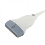
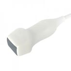
Linear Probes
A linear probe uses high-frequency ultrasound to create high-resolution images of structures near the body surface. This makes the probe ideal for vascular imaging and certain procedures such as central line placement.
Linear names get their name from the crystal arrangement being linear. The shape of the beam is rectangular, and the near-field resolution is good.
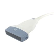
Convex Probes
Convex probes (also called curved linear probes) have a curved array that allows for a wider field of view at a lower frequency. The beam has a convex shape that makes the transducer ideal for deeper organ imaging examinations. Convex probes are primarily used for abdominal scans due to their wider depth and deeper penetration.
 Endocavitary Probes
Endocavitary probes are used less frequently in the ED but do afford advantages for specific studies. The wand-shaped design of this type of probe allows for examination of body cavities such as the oropharynx, female pelvic organs, and the male prostate.
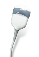
Phased Array/Cardiac Probes
Phased array or cardiac probes have a smaller handle with a square-shaped lens and array. Usually, they scan images of the heart. Phased array probes will have greater depth to reach the heart and produce an image.
%20Probes.png?width=189&height=142&name=Transesophageal%20(TEE)%20Probes.png)
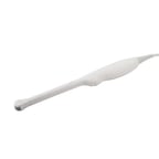
Transesophageal (TEE) Probes
TEE probes are cardiac-type probes that provide an obstructed image of the heart by inserting the probe into the esophagus, and into the patient’s stomach. These probes move in 4 different directions, and movement is controlled by the handle controls.
3D/4D Probes
3D probes function the same as 2D probes, with the exception of a moving array. The array inside the probe moves in a sweeping motion and captures slices of images from different sides. The captured slices are then all put together to produce a 3D still image, or a 4D live image.
Choosing the Right Ultrasound Probe
So, now that you have the different types of probes at your disposal, how do you choose the right probe? There are a few variables that factor into choosing the correct ultrasound probe.
Frequency
The sound waves that come from ceramic crystals, called piezoelectrics, generate signals at a particular speed and distance, which are measured in MHz, or megahertz.
Higher frequency equals better resolution, lower frequency equals better penetration.
Smaller wavelengths are more easily reflected or refracted in the superficial tissues than longer wavelengths. As the wavelength is increased (or frequency decreased), the ultrasound will penetrate deeper. As the wavelength is decreased (or frequency is increased), the ultrasound beam will have a shallower penetration.
Depth
The Depth is a special knob for adjusting the distance of the field of view. Structures within the field of view can be moved far or closer by adjusting the Depth. Lower frequency allows for imaging of nerves and tissue that require deeper penetration.
Shape of Array
The grayish rubber material at the end of the probe is called the “array”. The array covers the crystal elements that produce the sound waves to create an image. The array determines the shape of the image you’ll see. For example, a flat array will show a rectangular image.
Length of Array
A longer array will fit more anatomy beneath it, making it more appropriate for larger or deeper structures. A shorter array is appropriate for smaller structures so they appear larger on the screen relative to the probe.
👉 Ultrasound Training for Physicians: Why Your Practice Needs Ultrasound
Learning Ultrasound with SonoSim
SonoSim has created a breakthrough method for learning and teaching ultrasound that is now well proven. SonoSim Ultrasound Training overcomes longstanding barriers to ultrasound education. It complements clinical training by providing remote hands-on ultrasound training, didactic instruction, and assessment.
The SonoSim Ultrasound Training Solution is a software program that teaches healthcare providers how to perform and interpret ultrasound, particularly in a point-of-care setting. Specifically, the platform is delivered in a modular format comprised of online courses coupled with mastery questions, and an innovative laptop-based ultrasound simulator. Only SonoSim allows you to scan ultrasound cases obtained from real patients in a safe, simulated environment, anytime, anywhere.
CTA Link