


-
SPI Test Prep
-
OB/GYN Test Prep
-
QuestionBank
Sonography Principles & Instrumentation (SPI) Exam Preparation
With over 1,500 peer-reviewed questions mapped to the latest ARDMS SPI test requirements, interactive quizzes, and timed mock exams, SonoSim helps learners prepare with confidence. Just over 250,000 quizzes and mastery tests have already been completed, and students using SPI Test Prep have 4x better odds of passing the SPI exam on their first try, strengthening both their knowledge and clinical readiness.
Obstetrics & Gynecology (OB/GYN) Exam Preparation
SonoSim provides 1,350 peer-reviewed questions, complete with multimedia explainers and mapped to the ARDMS OB/GYN exam. Plus, users get unlimited weighted mock exams. Just another way SonoSim helps learners prepare with confidence. Each question features a single best answer with a detailed rationale and explanations for incorrect options, while a mix of text-, image-, and video-based questions reinforces the interpretation skills learners need to recognize pathology rather than just memorize answers.

Self-Test. Anytime. Anywhere.
With SonoSim’s 6,100-question and growing QuestionBank, learners can access a variety of on-demand, multimedia, short practice quizzes that can be completed in minutes. Quiz takers receive real-time expert feedback upon answering each question, perfect for quick study sessions and test preparation. The growing SonoSim QuestionBank is accessible on any device.


%20Examination.png?width=175&height=139&name=Abdomen(AB)%20Examination.png "Ultrasound image depicting abdomen content for diagnostic medical sonography programs")
%20Examination.png?width=175&height=139&name=Adult%20Echocardiography%20(AE)%20Examination.png "Ultrasound sound image of the heart showing SonoSim's echocardiography cases for DMS on demand")
%20Examination.png?width=175&height=139&name=Breast%20(BR)%20Examination.png "Ultrasound image of SonoSim's breast ultrasound content")
%20Examination.png?width=175&height=139&name=Musculoskeletal%20Sonographer%20(MSKS)%20Examination.png "MSK ultrasound image depicting SonoSim's MSK DMS on demand system")
%20Examination.png?width=175&height=139&name=Pediatric%20Sonography%20(PS)%20Examination.png "Pediatric sonography image for diagnostic medical sonography training")
%20Examination.png?width=175&height=139&name=Vascular%20Technology%20(VT)%20Examination.png "Vascular technology ultrasound image for training DMS on demand")
%20Examination.png?width=175&height=139&name=Obstetrics%20%26%20Gynecology%20(OBGYN)%20Examination.png "Obstetric ultrasound image to represent SonoSim modules that cover obstetrics and gynecology for DMS")


Breast Anatomy & Physiology Ultrasound Training
Course Highlights |
SonoSimulator Scanning Cases |
|
The course in this module covers topics including:
|
The 3 real patient scanning cases in this module include and cover:
|
Focused Cardiac Ultrasound (FoCUS) – Part I: Advanced Clinical Module
Course Highlights |
SonoSimulator Scanning Cases |
|
The course in this module covers topics including:
|
The 10 real patient scanning cases in this module include and cover:
|
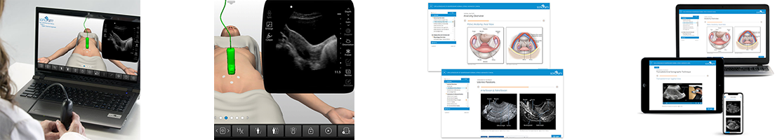
GYN Ultrasound of Nonpregnant Normal Uterus Advanced Clinical Training
Course Highlights |
SonoSimulator Scanning Cases |
|
The course in this module covers topics including:
|
The 10 real patient scanning cases in this module include and cover:
|
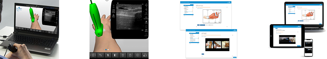
Musculoskeletal: Core Clinical Module
Course Highlights |
SonoSimulator Scanning Cases |
|
The course in this module covers topics including:
|
The 10 real patient scanning cases in this module include and cover:
|
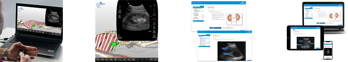
Renal: Core Clinical Module
Course Highlights |
SonoSimulator Scanning Cases |
|
The course in this module covers topics including:
|
The 10 real patient scanning cases in this module include and cover:
|


