🗞️ Ultrasound Application News
The use of Point-of-care ultrasound (POCUS) to improve patient care continues to expand. A recent study reveals how ICU triage teams are using POCUS to evaluate and manage deteriorating patients in hospital wards, improving diagnostic accuracy and guiding critical interventions. Discover how this versatile tool is transforming care in Internal Medicine, Family Medicine, Emergency Medicine, and beyond. Dive into the full story on our blog.
📚 Case Study: Cardiology
Take a look at this ultrasound case study!
Background:
- The patient has a prior history of a pacemaker placement and a Mitraclip™
- The patient undergoes an echocardiographic examination
👇 What type of pathology is in this clip?
👩⚕️ Scanning & Imaging Tips: Doppler Imaging Artifacts - Signal Aliasing
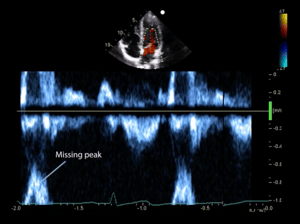
Signal aliasing is a common Doppler artifact caused when high-velocity blood flow exceeds the Nyquist Limit, often seen in valvular pathologies like stenosis or regurgitation. This results in spectral Doppler displaying flow as moving both toward and away from the transducer or waveforms being "cut off" and reappearing on the opposite baseline side.
To address aliasing:
- Shift the spectral Doppler baseline to adjust the graph displayed.
- Increase the velocity scale to enhance the sampling rate.
- Use continuous-wave Doppler to capture higher velocities accurately.
Understanding and managing signal aliasing ensures accurate interpretation of Doppler findings.

🔎 Scan & Seek - Fun with Physics
Keep watching below to see what the unusual subject we scanned this month was!
.gif?width=680&height=507&name=TSW%20-%20December%202024%20Media%20(2).gif)
This scan of a lettuce in a water bath demonstrates water trapped between the layers of lettuce leaves. Water acts as a great conductor for sound waves, allowing them to pass through with minimal resistance. The result? Acoustic shadowing! The area behind the fluid appears brighter than surrounding tissues—just like in this image, where the left side lights up more significantly! ✨ This effect helps us understand how areas posterior to fluid-filled structures (like cysts or the bladder) appear brighter on ultrasound due to the low impedance of water. Ultrasound physics at work—right in your veggie drawer! 🌱
🧠 Challenge Case of the Month: Obstetrics
Case History: This 32-year-old G1P0 female presents to the emergency department with reports of being pregnant. She states that her LMP was approximately 11 weeks ago, and reports pelvic cramping and vaginal bleeding for one day.
View this transabdominal ultrasound and evaluate the uterus.
What are the relevant sonographic findings?👇
💡 SonoSim Tips & Tricks
Unlock Ultrasound Knowledge with QuestionBank!
SonoSim’s QuestionBank is a powerful, standalone resource designed to help you test and refine your ultrasound knowledge anytime, anywhere. It provides an alternative learning method using Q&A sets and case-based scenarios.
How does QuestionBank Complement Knowledge Checks and Mastery Tests?
- Knowledge Checks: In the courses, these reinforce the lesson concepts
- Mastery Tests: Summary assessments document course mastery.
- QuestionBank: A comprehensive collection of Q&A sets and case-based scenarios that map to specific ultrasound applications.
With QuestionBank, you can strengthen core concepts, apply knowledge to real-world scenarios, and study on your schedule—all from your phone, tablet, or computer. Explore this invaluable tool today and take your ultrasound training to the next level!
😂 LOLtrasound

Image Credit: FX Production
📲 Follow Us on Social!

