🙌 It's National Critical Care Awareness & Recognition Month (NCCARM)
We celebrate the unwavering and inspiring commitment of critical care providers worldwide. In honor of this important month for healthcare, this issue is all about Critical Care ultrasound education & information. #CritCareMonth
🗞️ Ultrasound Application News
Discover how POCUS enhances patient care beyond the ICU in our new blog post! A new study showcasing the impact of POCUS by ICU triage teams in general hospital wards is discussed.
Did you miss our blog post on the use of Nurse-Performed Point-of-Care Ultrasound (NP-POCUS) in the management of septic patients in emergency departments?
💥 SonoSim Updates
We're excited to share some highlights from our May release, which brings valuable improvements to the platform. Check out what's new:
- Advanced Content Search: Our reimagined search engine helps you find the content you're looking for with one click. Case Studies have recently been added to the search results.
- Doppler and Caliper Enhancements: Improved precision for Doppler video measurements, increased video player resolution, and optimized playback controls for easier measurement readings.
Dive into the blog post to learn more about these updates and how they can enhance your learning experience!
📚 Case Study: Large Pericardial Effusion
Take a look at this ultrasound case study!

Background:
The patient is awake and alert. He is not in visible distress and does not recall what happened.
- BP: 112/90 mmHg
- HR: 52 bpm
- RR: 18 bpm
- O2: 96% on RA
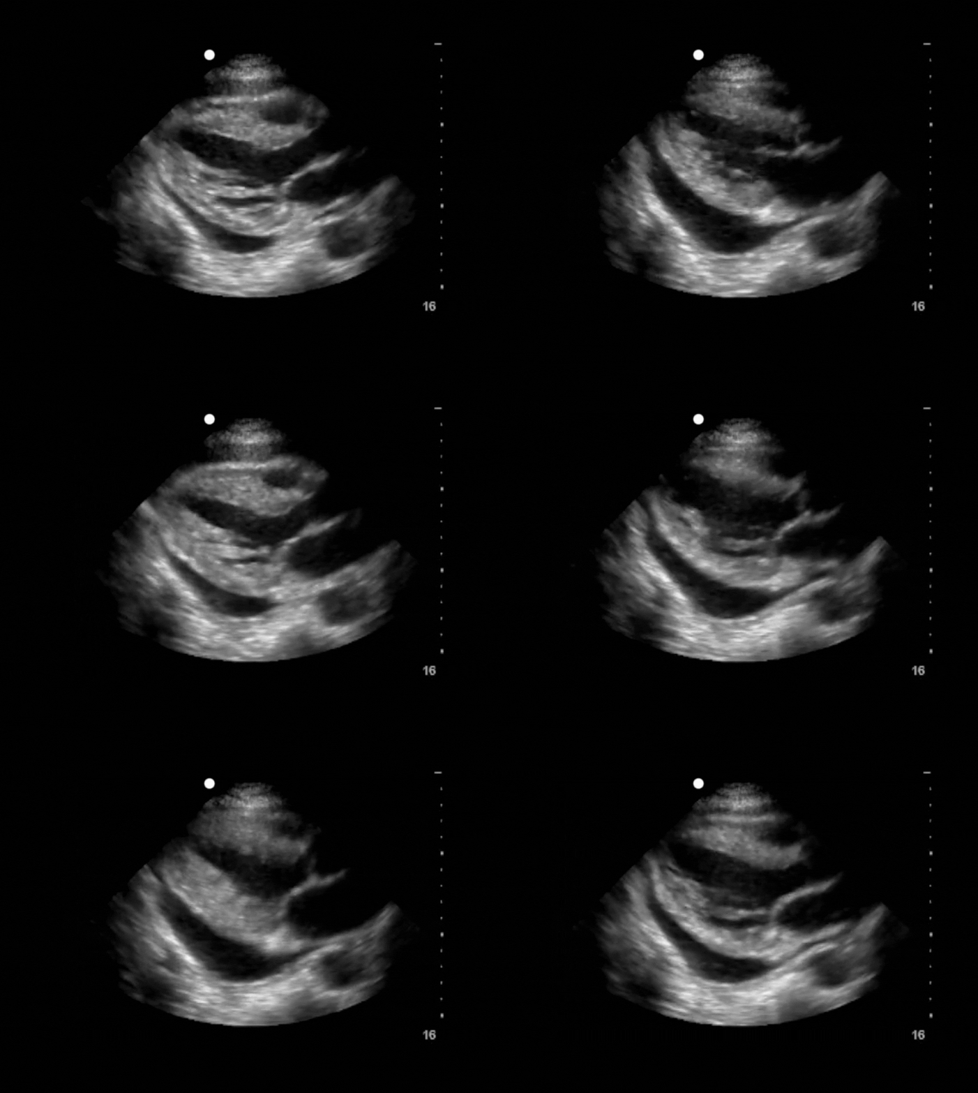
A large circumferential pericardial effusion is seen in this parasternal long-axis view of the heart. A "swinging heart sign" is evident and often corresponds with an electrical alternans pattern on the patient’s EKG. There is a subtle diastolic collapse of the free wall of the right ventricle during several frames of this clip.
Both findings are supportive of cardiac tamponade physiology, but remember cardiac tamponade is a clinical diagnosis. Given the patient’s current status obtaining additional clinical history, physical findings, and additional cardiac views and parameters (e.g., IVC inlet view, MV/TV inflow velocities) is necessary to characterize the physiologic impact of this large pericardial effusion.
👩⚕️ Scanning & Imaging Tips: Abdominal aorta imaging tips

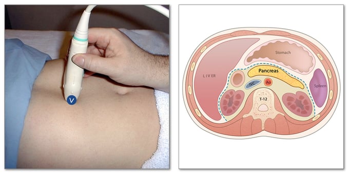
When performing abdominal ultrasound, use the spine shadow as a primary landmark to identify the aorta and adjacent structures. Press with both hands to depress bowel loops for a clearer view. The aorta is just above the spine shadow, with the IVC to the right. Look for the superior mesenteric artery (SMA) anterior to the aorta, surrounded by hyperechoic tissue, and the splenic vein crossing over the SMA toward the liver. The pancreas lies above the splenic vein, and the less echogenic liver is the most anterior structure. Identify these landmarks to enhance your scanning accuracy.
🔎 Scan & Seek
Keep watching below to see what the unusual subject we scanned this month was!
.gif?width=680&height=507&name=TSW%20-%20May%202024%20Media%20(1).gif)
Surprise! The ultrasound revealed that our mystery object is a flower. We see distinct outer petals, while the center appears hazy due to the trapped air, which scatters and reflects the sound waves in different directions.
🧠 Challenge Case of the Month: Abdomen Case 9
Case History: This 84-year-old male presents with syncope, abdominal pain, and hypotension.
See the ultrasound below and evaluate his mid-aorta. 👇
SonoSim Members: Don't forget that you get regular additions to Challenge Cases. Check them out!
💡 SonoSim Tips & Tricks
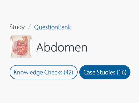
Have you noticed the two buttons below each topic area in QuestionBank?
- Case Studies: Real case-based scenarios and ultrasound clips enhance knowledge & decision-making skills
- Knowledge Checks: Engaging quiz-based format, across a broad range of topics, improves ultrasound image interpretation.
Toggle between these filters to streamline your study experience!

😂 LOLtrasound

📲 Follow Us on Social!

