🗞️ Ultrasound Application News
Check out these 3 recent blog posts to help you stay ahead in the world of medical imaging and patient care:
Explore how early ultrasound education enhances medical students' visual-spatial skills and understanding of cross-sectional imaging. These abilities are crucial for interpreting complex anatomical relationships, benefiting fields like radiology and surgery. Dive into the study's key findings and its impact on medical education in our recent blog post.
In another blog post, we address the ongoing sonographer shortage and its implications for healthcare. As demand for ultrasound exams outpaces the supply of trained professionals, this shortage challenges patient care and facility operations. Discover the contributing factors and explore potential solutions to ensure the continued delivery of quality ultrasound services.
Finally, we highlight the growing importance of point-of-care ultrasound (POCUS) for hospitalists. This essential tool improves diagnostic accuracy, speeds patient care, and increases procedural safety. Learn why POCUS is recommended as a must-have skill in hospital medicine and how it’s transforming care in our latest blog post.
📚 Case Study: Hit and Run
Take a look at this ultrasound case study!
Background:
- 50 yo male presents to the ED in moderate distress with diffuse abdominal pain one day after being struck by a car.
- Vitals: BP = 90/40 mmHg; HR = 104 bpm, RR = 20 bpm; O₂ sat = 94% on room air
Which of the following choices best describes the accompanying sonographic findings?
Isolated pocket of free fluid within Morison’s pouch.
- Diffuse anechoic fluid collection seen in RUQ, LUQ, and Suprapubic windows.
- Large-volume ascites associated in a setting of liver cirrhosis and associated portal hypertension.
Get the answer by watching the clip below!
📚 Case Study: Left Leg Swelling
Background:
- 16yo male with cyanotic congenital heart disease presents with left leg swelling for several days
- Left leg is edematous from the proximal thigh downward, but displays normal color, 2+ palpable pulses, and normal capillary refill.
Which of the following statements best characterizes the sonographic findings in the accompanying ultrasound clip taken along the proximal left thigh of this patient?
- Reduced compressibility in the external iliac vein
- Reduced compressibility in the left common femoral vein
- Reduced compressibility in the left great saphenous vein
- Reduced compressibility in the left small saphenous vein
Get the answer by watching the clip below!
👩⚕️ Scanning & Imaging Tips: OB Six-Step Examination
The OB Six-Step Examination Protocol is a vital tool that enhances maternal care. By quickly assessing fetal presentation, cardiac activity, amniotic fluid levels, and more, this structured approach helps healthcare providers make timely decisions, reducing the risk of complications during delivery. In resource-limited settings, where every minute counts, this protocol can be life-saving—playing a key role in reducing maternal mortality rates by ensuring early detection of critical conditions.

👩⚕️ Scanning & Imaging Tips: Overcoming Gassed-Out GI Images

While most patients are initially scanned in a supine body position, movement to a semi-upright, lateral oblique, or lateral decubitus position may be useful.
Do not be deterred when initially placing the transducer on the abdomen yields a “gassed-out” image. Adjusting patient position may help to displace air-filled bowel loops that are degrading image quality.
Using graded compression may also improve ultrasound image quality. Graded compression is applied to the transducer to displace gas in the bowel loops located beneath the transducer (Puylaert). Gentle, but continuous pressure should be used to avoid causing pain, which could cause reflex contraction of the abdominal wall musculature. If the examiner is evaluating a painful condition, analgesic medication can minimize pressure-induced discomfort. Having the patient fast or only drink fluids are alternative methods of decreasing gas artifacts and improving image quality (Folvik et al.).
🔎 Scan & Seek - Fun with Physics
Keep watching below to see what the unusual subject we scanned this month was!
.gif?width=680&height=507&name=TSW%20-%20October%202024%20Media%20(3).gif)
When you scan a pumpkin, you can see some interesting physics at play! The pumpkin’s thick, dense shell creates high attenuation, which means the ultrasound waves lose energy as they pass through it. As a result, less sound energy reaches deeper tissues, causing a posterior acoustic shadow—a dark area behind the shell where fewer echoes are returned. This is similar to how bones or calcifications appear in human ultrasound scans!
🧠 Challenge Case of the Month: Abdomen
Case History: This 17-year-old patient with a history of hemophagocytic lymphohistiocytosis (HLH) and liver cirrhosis complicated by portal hypertension, chronic portal vein thrombosis with cavernous transformation, and esophageal varices that was treated with a transjugular intrahepatic portosystemic shunt (TIPS) now presents to the ED with lower abdominal pain and melena. She is hemodynamically stable.
Click to watch the clip and evaluate her TIPS patency.👇
🧠 Challenge Case of the Month: Head/Neck
Case History: This 58-year-old female presents with painless anterior neck swelling thought to represent an enlarged thyroid gland.
Click to watch the clip and assess her thyroid.👇
🔑 SonoSim Members: Don't forget that you have hundreds of Challenge Cases and that you get regular additions. Check them out in the SonoSimulator Case Library!
💥 SonoSim Updates
We’re thrilled to announce several new updates to enhance the SonoSim ultrasound learning experience!
New in My Image Portfolio: Edit Annotations & Delete Images
Now available to all users! You can edit annotations and delete images directly within your My Image Portfolio on the web, making it easier to manage, organize, & correct your ultrasound images for study, review, and sharing.
Coming Soon: Gastric Ultrasound
Enhance your ability to assess gastric contents and volume with POCUS. Learn essential skills for evaluating gastric emptying and aspiration risk during procedural sedation and anesthesia. This tool is crucial for preoperative assessments of high-risk patients, or where fasting protocols may not apply.
These updates are designed to support your ongoing journey to ultrasound mastery. Stay tuned for more!
💡 SonoSim Tips & Tricks
SonoSim’s Automated Image Assessment (AIA) instantly evaluates ultrasound images, comparing them to optimal scans from professional sonographers, and providing immediate feedback—saving instructors valuable time. Please note, AIA is currently available only for Anatomy & Physiology scanning cases.
To activate AIA, head to My SonoSim Dashboard > Track > Image Feedback and flip the switch in the top right corner!
.gif?width=527&height=290&name=TSW---October-2024-Media-(3).gif)
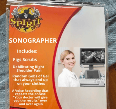
😂 LOLtrasound

Credit: @thesonographerlife
📲 Follow Us on Social!

