2025
June 2025 - Protocol Post
Highlighting advancements in POCUS Protocols, this June 2025 issue of SonoSim Wave explores emerging trends, tools, and clinical applications in...
Discover MSK ultrasound insights in this July 2025 issue—case studies, dynamic scanning tips, imaging tricks, and updates from SonoSim.
🗞️ Ultrasound Application News
A study in the American Journal of Emergency Medicine introduces the lateral approach water bath, a novel ultrasound technique that significantly improves hand imaging. By stabilizing the probe against the side of a container rather than suspending it in water, clinicians gain better image quality, reduced motion artifact, and improved patient comfort. This technique shows promise for enhancing MSK ultrasound in sports medicine, orthopedics, and emergency care. Read the full blog post to explore the findings!
📚 Case Study: Ankle
What are the relevant sonographic findings?
Background:
👩⚕️ Scanning & Imaging Tips: Dynamic Scanning
One of the most impressive aspects of musculoskeletal ultrasound is its ability to observe dynamic anatomy and various relationships, as well as view structures in their full range of motion. In the above clip, the flexor tendons can be observed moving in a short-axis view. Similarly, the extensor tendons can be examined to track movement and identify individual tendons. Tendon movement is better appreciated in a long-axis view in the second portion of this ultrasound video.
Pro Tip: Always compare with the contralateral side for subtle asymmetries.
🔎 Scan & Seek - Fun with Physics
Look below to see the flamingo we hid in this ultrasound image of the volar plate.
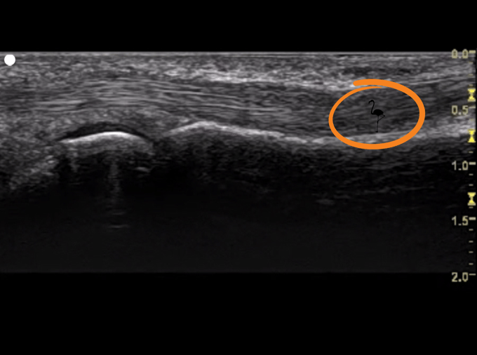
The volar plate is a fibrocartilaginous structure seen overlying the joint, arising from the base of the distal bone, coursing proximally. It overlies the volar aspect of the digital joints, including the metacarpophalangeal, proximal interphalangeal, and distal interphalangeal joints. The volar plate’s shape and appearance is similar at each of these locations. It has a homogeneous, echogenic texture.
Here, one sees the third digit volar plate overlying the metacarpophalangeal joint. Along the base of the proximal phalanx, it crosses the joint space proximally. The anechoic rim between the metacarpal head and the volar plate is the articular cartilage. Notice the flexor tendons, which lie superficial to the volar plate. In the near field, just beneath the transducer, subcutaneous fat and connective tissue outline the tendons.
🧠 Challenge Case of the Month: MSK
Case History: This 20-year-old male presents with a history of falling while jogging and now has a foreshortened left leg and deformed right forearm.
Examine his left femur with ultrasonography.👇
✨ SonoSim Updates
A New & Improved Course Experience - Rolling Out Soon!
We’re making it easier than ever to navigate and complete your SonoSim courses. Our upcoming New Course-Taking Experience brings clearer course outlines, improved progress tracking, and a more intuitive interface—designed to help you learn more efficiently.
This update will be introduced gradually on a rolling basis, starting in August, to ensure a smooth transition for all members. When it becomes available to you, we’ll provide guidance on what to expect and how to opt in. Stay tuned!
💡 SonoSim Tips & Tricks
New to scanning with the SonoSimulator®?
This quick orientation walks you through everything you need to know to get started: how to hold and calibrate the probe, scan cases, and use key features like virtual controls, case guidance, and assignments. In just 5–10 minutes, you'll build confidence in navigating the Scan element of the SonoSim Ecosystem, so you can focus on mastering ultrasound, not troubleshooting tech.
SonoSim continues to support our members not only with the easiest way to learn and teach ultrasonography, but also with comprehensive orientation and onboarding to the ultrasound education ecosystem.
😂 LOLtrasound
.png?width=466&height=466&name=Ultrasound%20Memes%20(5).png)
IB: @thebluntsonographer
📲 Follow Us on Social!

.png?width=600&height=314&name=TSW%20-%20Closing%20Ad%20(1).png)
Highlighting advancements in POCUS Protocols, this June 2025 issue of SonoSim Wave explores emerging trends, tools, and clinical applications in...
Highlighting advancements in critical care ultrasound & ultrasound-guided procedures.
Discover unique ultrasound insights in this August 2025 issue—ocular, soft tissue, foreign body localization, and pediatric MSK cases, plus expert...