2025
August 2025 - Untapped Ultrasound
Discover unique ultrasound insights in this August 2025 issue—ocular, soft tissue, foreign body localization, and pediatric MSK cases, plus expert...
Discover unique ultrasound insights in this September 2025 issue—OB-GYN case study, Doppler optimization tips, scanning challenge cases, and expert updates from SonoSim.
🗞️ Ultrasound Application News
Can Sonographers Reliably Score Thyroid Nodules Using TI-RADS? A new study suggests they can. Researchers found trained sonographers achieved comparable, and in some cases superior, accuracy to radiologists when scoring thyroid nodules with TI-RADS. These findings highlight a potential pathway to improve diagnostic efficiency and resource allocation in thyroid imaging. Read the full blog post for more.
What an incredible time we had connecting with sonography educators and learners at SDMS 2025 in Denver! 🎉 Thank you to everyone who stopped by Booth #212 to experience how SonoSim is helping programs build a complete ultrasound education ecosystem.
📌 Couldn’t make it, or want to revisit what we shared?

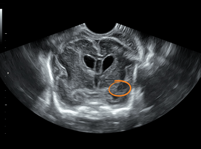
📚 Case Study: OB/GYN
Background:
This 40-year-old female with a history of prior bilateral tubal ligation is referred to the ED for a positive pregnancy test noted during a routine obstetrics and gynecology (OB/GYN) appointment. She undergoes a pelvic ultrasound exam.
Which of the following choices best describes the sonographic findings seen in the accompanying ultrasound clips?
A. Hemorrhagic cyst
B. Endometrioma
C. Intrauterine pregnancy
D. Ectopic pregnancy
Correct Answer: D. Ectopic pregnancy
👩⚕️ Scanning & Imaging Tips: Optimizing Signal Aliasing
Please examine this accompanying ultrasound image of a blood vessel that was obtained using color Doppler.
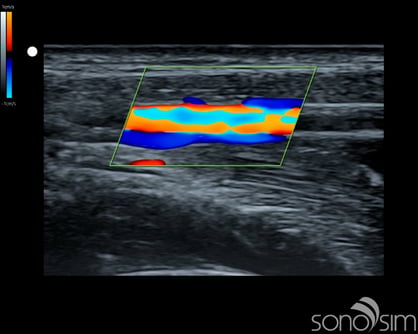
Which of the following interventions will best optimize visualization of blood flow within the Doppler color box?
A. Shift baseline upward
B. Decrease color Doppler box size
C. Decrease pulse repetition frequency (PRF)
D. Increase velocity scale
The correct choice is increase velocity scale. The example image appears to show multi-directional flow at the center of the vessel. However, it also shows relatively laminar flow (smooth, layered flow) without turbulent mixing. This indicates an aliasing artifact in which the velocity exceeds the Nyquist limit. Note that the directional change also occurs in the center of the vessel where velocity would be expected to be highest. The adjacent colors within the vessel (orange next to blue-aqua) are located along polar opposites of the color map, which further indicates this is signal aliasing artifact, as one would expect the dark adjacent colors across the black base-line within the vessel to be located adjacent to each other on the color map with bidirectional flow. Increasing the velocity scale will alleviate this artifact. Decreasing the PRF will reduce the Nyquist limit (creating more aliasing), while changing the box size will only change the frame rate.
🔎 Scan & Seek - Fun with Physics
Look below to see the leaf we hid in this ultrasound image.
🧠 Challenge Case of the Month: Abdomen
Case History: This 18 year-old-male with history of IBD presents with abdominal pain and distention.
Please evaluate his abdomen with ultrasound.👇
✨ SonoSim Updates
Check out all that’s new in September!
💡 SonoSim Tips & Tricks

Want to get the most out of SonoSim’s Study tools?
This quick orientation will guide you through the Study Element of the SonoSim® Ecosystem, helping you build a smarter study strategy in just 5-10 minutes. You’ll get a clear overview of all the SonoSim Study Tools available, explore Challenge Cases within the SonoSimulator®, and learn how to test and reinforce your knowledge with SonoSim's QuestionBank and Test Prep tools.
Learners preparing for the ARDMS SPI or a specialty certification exam and POCUS learners preparing FPD-AEMUS or the CCEeXAM, SonoSim Study Tools can support and enhance learning efforts.
😂 LOLtrasound
Credit: @sonogiggles
📲 Follow Us on Social!

.png?width=600&height=314&name=TSW%20-%20Closing%20Ad%20(3).png)
Discover unique ultrasound insights in this August 2025 issue—ocular, soft tissue, foreign body localization, and pediatric MSK cases, plus expert...
The September 2024 issue of the SonoSim Wave explores joint and MSK ultrasound applications and use cases.
Explore the January 2025 issue of SonoSim Wave, featuring advancements in ultrasound applications, case studies, expert tips, and software updates to...