2025
November 2025 - Movember Monthly
Discover the November 2025 SonoSim Wave with Movember highlights, men’s health insights, and ultrasound education updates for instructors and...
The October 2026 SonoSim Wave marks Medical Ultrasound Awareness Month with neonatal lung ultrasound, ovarian torsion tips, and the new Assignments feature.
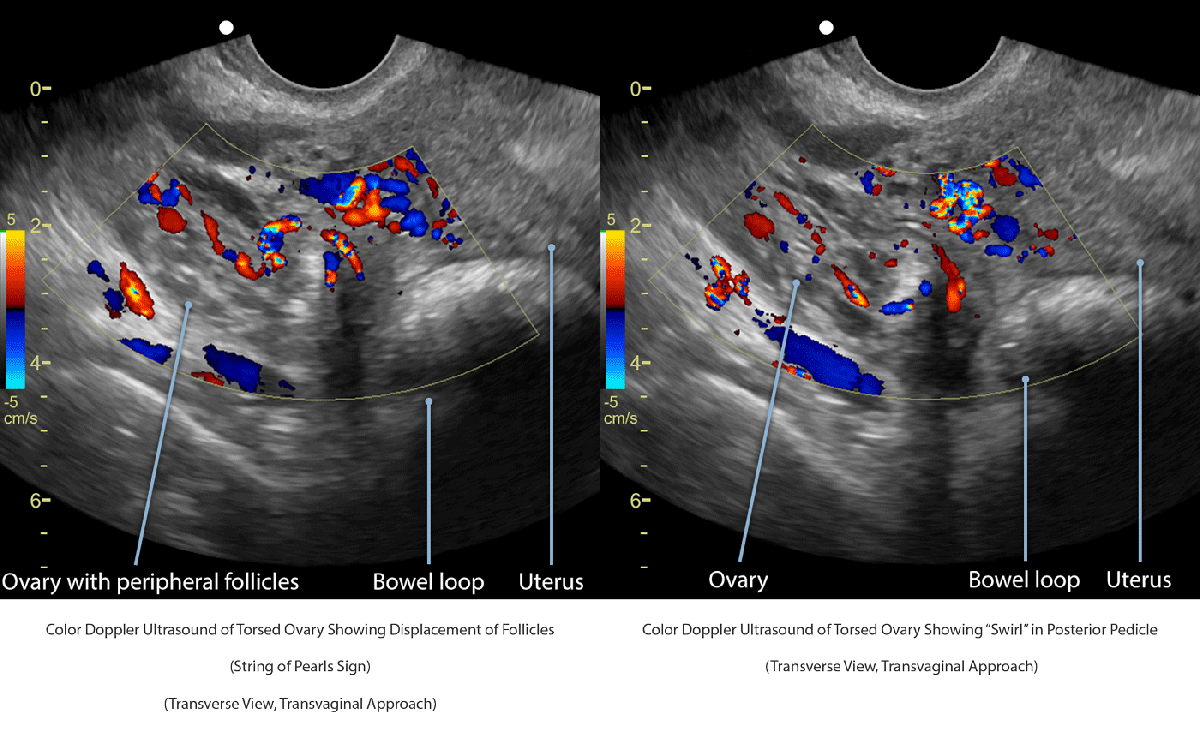
Adnexal torsion may cause ovarian displacement, enlargement, edema, and peripheral follicle displacement, known as the “string of pearls sign” (Chang et al.). The accompanying color Doppler transvaginal ultrasound images demonstrate a torsed right ovary with peripheral follicles and a twisted vascular pedicle, known as the “whirlpool sign.”
Color and pulsed-wave Doppler findings vary with the degree of vascular compromise and may demonstrate reduced or absent arterial and venous flow (Shadinger et al.). Importantly, normal Doppler flow does not exclude ovarian torsion, as the ovary’s dual blood supply or intermittent or partial torsion may preserve arterial perfusion.
Check out GYN Ultrasound Nonmalignant Adnexal Conditions Advanced Clinical
Albayram F, Hamper UM. Ovarian and adnexal torsion: spectrum of sonographic findings with pathologic correlation. J Ultrasound Med. 2001 Oct;20(10):1083-1089.
Chang HC, Bhatt S, Dogra VS. Pearls and pitfalls in diagnosis of ovarian torsion. Radiographics. 2008 Sep-Oct;28(5):1355-1368.
Pena JE, Ufberg D, Cooney N, et al. Usefulness of Doppler sonography in the diagnosis of ovarian torsion. Fertil Steril 2000 May;73(5):1047-1050.
Servaes S, Zurakowski D, Laufer MR, et al. Sonographic findings of ovarian torsion in children. Pediatr Radiol. 2007 May;37(5):446-451.
Shadinger L, Andreotti R, Kurian R. Preoperative sonographic and clinical characteristics as predictors of ovarian torsion. J Ultrasound Med. 2008 Jan;27(1):7-13.
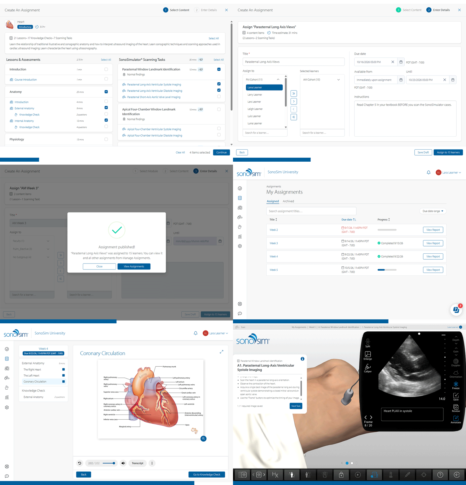
Coming November 2: We're excited to bring you In-Platform Assignments 🎉
A new feature designed to make assigning coursework, managing learning activities, and tracking progress easier for educators and learners is coming soon.
For Admins & Instructors: Create and customize assignments, assign specific modules or scanning cases to individuals or groups, and schedule coursework in advance. Easily manage active, scheduled, draft, and archived assignments, and track learner progress and completion with built-in reporting tools.
For Learners: Stay on top of your coursework with a dedicated 'My Assignments' section. View upcoming due dates, track completion progress, access assigned learning activities, and receive notifications when new assignments are available.
More details soon! 🎉
We want to hear from you!
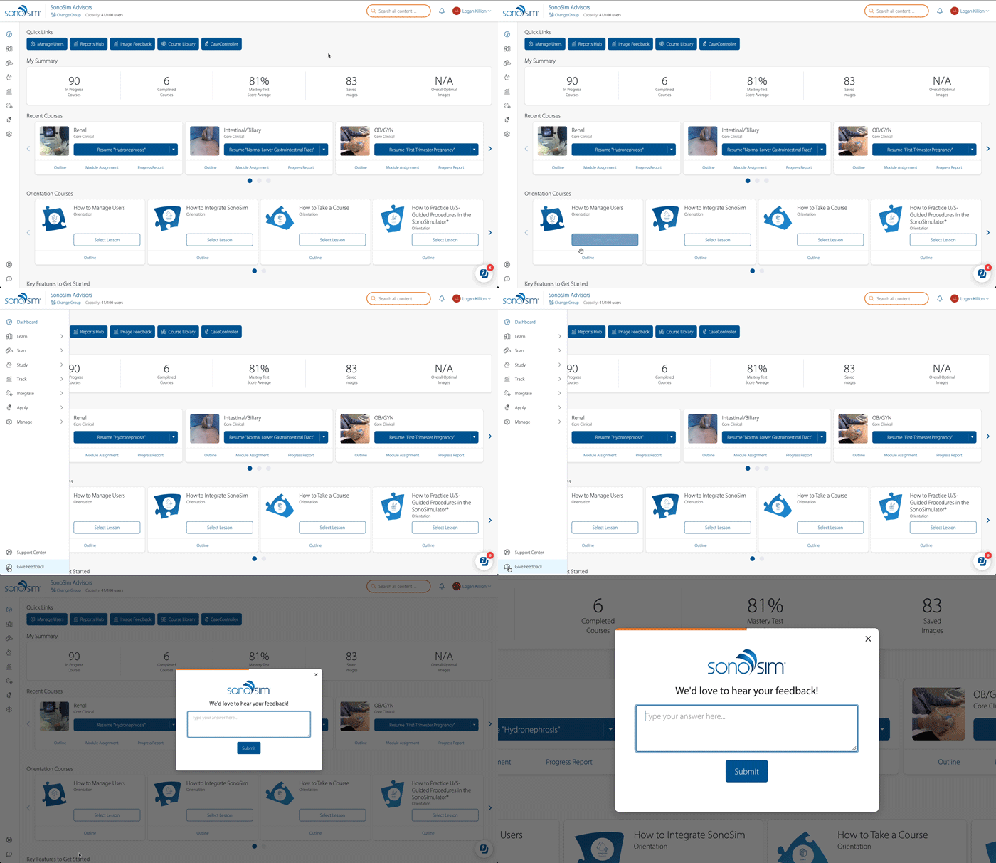
Your feedback helps us make SonoSim better. Whether you have a suggestion, an idea for improvement, or something you'd like to share, simply click the "Give Feedback" button within your SonoSim Dashboard. Every submission is read by a real person on our team, and we're always eager to hear what you think!

📲 Follow Us on Social!

Discover the November 2025 SonoSim Wave with Movember highlights, men’s health insights, and ultrasound education updates for instructors and...
Discover the December 2025 SonoSim Wave with gastric ultrasound safety guidance for GLP-1 patients, featured GI scanning tips, new case studies,...
Highlighting advancements in critical care ultrasound & ultrasound-guided procedures.