🗞️ Ultrasound Application News
Check out our latest blog post, "The Potential of POCUS in Gastrointestinal Procedures," to understand how gastric Point-of-Care Ultrasonography (POCUS) is improving pre-endoscopy patient evaluations and reducing aspiration risks. See how SonoSim's ultrasound training can enhance your skills in gastrointestinal procedures. Click to read and learn more about the practical benefits of POCUS in clinical settings.
📚 Case Study: Acute Cholecystitis
Take a look at this ultrasound case study!
What we see:
- Thickened anterior gallbladder wall (>3mm)
- Gallstones with posterior acoustic shadowing
- Pericholecystic free fluid
- Sonographic Murphy's sign
This is an example showing gallstone shadowing, a thickened gallbladder wall, and pericholecystic fluid. When the operator compressed the gallbladder, the patient reported pain. This illustrates a positive sonographic Murphy’s sign. These are the ultrasound characteristics of acute cholecystitis. A sonographic Murphy’s sign is defined by a patient experiencing pain and inspiratory effort arrest following pressure on an ultrasound probe positioned over the gallbladder.
👩⚕️ Scanning & Imaging Tips: Gallbladder
The gallbladder can be found in so many different locations and imaging planes. The operator will often need to fan, rotate, and angle the transducer.
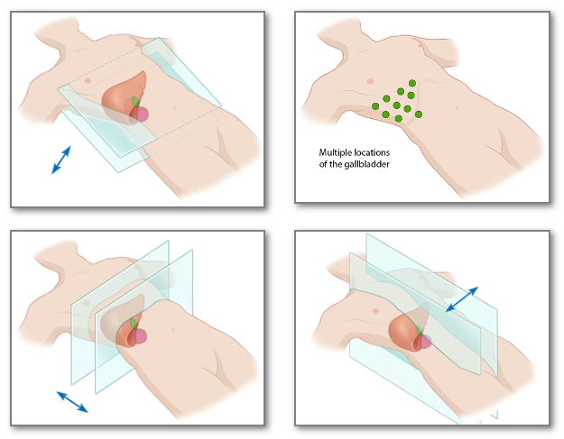
- The gallbladder is not aligned with standard sagittal and transverse body planes
- Use landmarks, like main lobar fissure, to find gallbladder
- Use different patient positions, breath holding, and multiple scanning approaches to improve views
- Use Doppler-mode imaging to differentiate between blood vessels and ducts
🔎 Scan & Seek
Keep watching below to see what the unusual subject we scanned this month was!
.gif?width=680&height=480&name=Scan%20%26%20Seek%20Reveals%20(1).gif)
🧠 Challenge Case of the Month: Abdomen Case 9
Case History: This 33-year-old male presents to the emergency department with right upper epigastric pain.
See the ultrasound below and evaluate his gallbladder.
SonoSim Members: Don't forget that you get regular additions to Challenge Cases. Check them out!
💥 SonoSim Updates
We're thrilled to bring you a bundle of fresh updates from April and a few extra you may have missed! Read the full release notes for more details.
- Orientation Courses:
- How to Integrate SonoSim: Designed to help you effectively use SonoSim's integration tools, learn to find ultrasound learning objectives specific to your industry, map them to relevant content, and assign guided scanning tasks.
- How to Practice U/S-Guided Procedures in the SonoSimulator®: Enhance your skills in ultrasound-guided procedures with our specially designed course. Practice crucial psychomotor and cognitive tasks in a risk-free environment.
- New QuestionBank Case Studies:
- Atrial Septal Defect & Ventricular Septal Defect
- Epididymitis
- Ectopic Pregnancy
- Internal Jugular Vein Thrombosis
- Retinal Detachment & Vitreous Hemorrhage
- New OB/GYN Challenge Cases:
- Fetal Duodenal Atresia
- Fetal Bilateral Hydronephrosis & Bladder with Keyhole Sign
- Umbilical Cord Mass
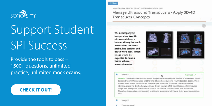
- SPI Test Prep: Our SPI Test Prep is receiving rave reviews! See for yourself. →
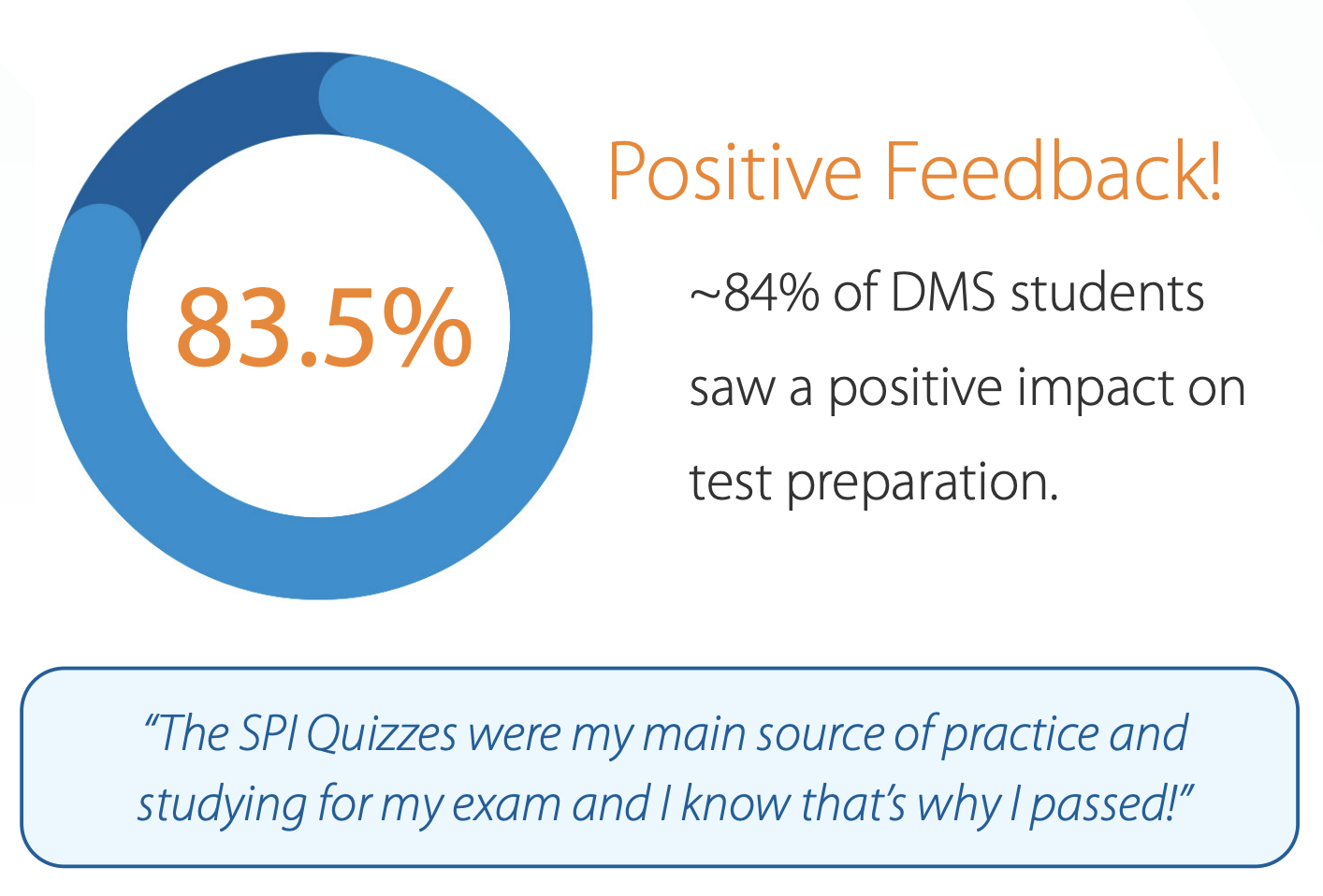
- Advanced Content Search: Our content search just got smarter! Find specific ultrasound topics and pathologies with ease and precision, thanks to our improved search functionality.
- SPI Individual Reports: Get detailed insights with our new report feature, offering individualized performance analytics for each learner.
For a complete overview of our recent releases and how they can enhance your ultrasound training, check out all of our release notes.
💡 SonoSim Tips & Tricks
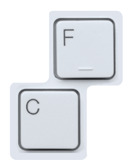
Take advantage of hot keys in the SonoSimulator®! Need to calibrate quickly? You can simply place your probe in the start position and hit the 'C' key to calibrate. Want to freeze your ultrasound image with ease? Hit the 'F' key while scanning. Happy scanning!
😂 LOLtrasound

📲 Follow Us on Social!

