
2026

November 2026 - Movember Memo

The SonoSim Wave Movember Memo covers prostate micro-ultrasound, a testicular trauma case, Doppler tips for torsion, and how to build a SonoSim Assignment.


🗞️ Ultrasound Application News

A randomized clinical trial published in JAMA explores the potential of high-resolution micro-ultrasound as an alternative to MRI-guided biopsy for prostate cancer diagnosis. The OPTIMUM trial found that micro-ultrasound-guided biopsy was noninferior to MRI fusion-guided biopsy in detecting clinically significant prostate cancer, highlighting its potential to improve imaging accessibility and streamline diagnostic workflows. Our latest blog breaks down the findings, their implications for prostate cancer detection, and the importance of ultrasound training in image-guided diagnosis.

You can read the full blog post here.


⭐ Special Feature: Men's Health Awareness Month 🎉

This November, we're joining the Movember movement to shine a spotlight on men's health! From raising awareness about prostate and testicular cancer to encouraging conversations around mental health and preventive care, this month is a reminder of the importance of prioritizing men's well-being. Here's to spreading awareness, starting conversations, and supporting healthier lives!


📚 Case of the Month

Background: This 21-year-old man presented with right hemiscrotal trauma following a motorcycle accident.

👇 View the findings video below for the diagnosis.


Check out Scrotum Anatomy & Physiology Ultrasound Training

🔑 SonoSim Members - Login to go to Scrotum Anatomy & Physiology Ultrasound Training


👩‍⚕️ Scanning & Imaging Tips: Pulsed-Wave Doppler in Testicular Ultrasound


Early in testicular torsion, the low-pressure venous system loses blood before higher-pressure arterial flow. Therefore, documenting both venous and arterial flow is key to addressing concerns of interrupted blood flow. Pulsed-wave Doppler is capable of distinguishing between arterial and venous flow patterns. Pulsed-wave Doppler should be set for low volume and slow flow to evaluate testicular arteries and veins.

Pulsed-wave testicular flow should be sampled in multiple locations. This is performed in combination with color or power Doppler, which shows where to assess for flow. As seen in this video, the color Doppler running in the background shows the operator where to position the pulsed-wave gate. The gate is moved around the testicle and tracings are obtained in various locations diffusely throughout the testicle, which mitigates sampling error. Both arterial and venous tracings should be documented.

Check out Scrotum Anatomy & Physiology Ultrasound Training

🔑 SonoSim Members - Login to go to Scrotum Anatomy & Physiology Ultrasound Training

🔎 Under the Probe

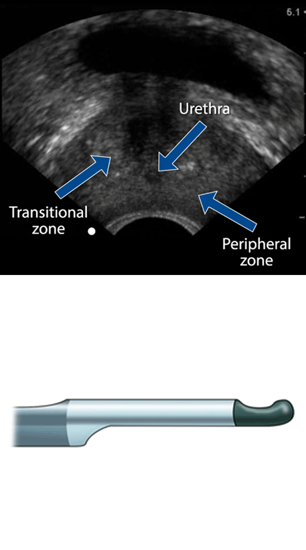
Which of the following statements best characterizes sonographic examination of the prostate gland?

Endocavitary ultrasound transducer, and a transverse ultrasound image of the prostate labeling the urethra, transitional zone, and peripheral zone

A. The prostate gland is best imaged with transabdominal sonography.

B. The prostate gland is best imaged with endorectal sonography.

C. An empty bladder facilitates transabdominal visualization of the prostate gland.

Read the explanation below for an in-depth overview.

The prostate gland is best imaged with endorectal sonography. However, one may be able to assess the overall size of the prostate gland using a transabdominal approach. The prostate is seen deep to the bladder and is best visualized when the bladder is urine-filled.


✨ SonoSim Updates

🎉 SonoSim Assignments Is Now Live!

We're excited to announce that SonoSim Assignments is officially live! This new in-platform feature makes it easier for educators to organize coursework, manage learning activities, and track progress, all within SonoSim.

For Admins & Instructors: Create customized assignments by selecting specific course lessons, knowledge checks, mastery tests, and scanning activities. Assign work to individuals or groups, set due dates, schedule coursework in advance, and track completion with assignment-specific reports. You can even share assignments with colleagues and copy existing ones for future use!

For Learners: Stay organized with the dedicated My Assignments section, where you can access coursework, view upcoming deadlines, track completion progress, and complete assigned scanning activities using the SonoSimulator®.

Ready to explore? Create your first assignment or check out our Release Notes to discover everything this exciting new feature has to offer!


💡 SonoSim Tips & Tricks

How to Build Your First Assignment

Ready to put the new SonoSim Assignments feature to work? Creating an assignment is easy! Head to Assignments > Manage Assignments in your MySonoSim Dashboard and click Create New Assignment. Choose a module, then customize your assignment by selecting specific lessons, knowledge checks, mastery tests, or scanning activities. Next, select your learners, add a title and optional instructions, and set your due dates. Once you're ready, publish your assignment, schedule it for later, or save it as a draft. It's that simple!


😂 LOLtrasound

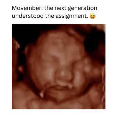
Movember wave meme 3 (2)


📲 Follow Us on Social!

Screenshot 2024-04-16 at 11.04.01 AM

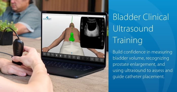
Bladder Ultrasound Ad_Final  

Similar posts

The SonoSim Wave
An Ultrasound Insights Newsletter

Get the latest trends in ultrasound training, education, & applications delivered to your inbox.

Previous editions