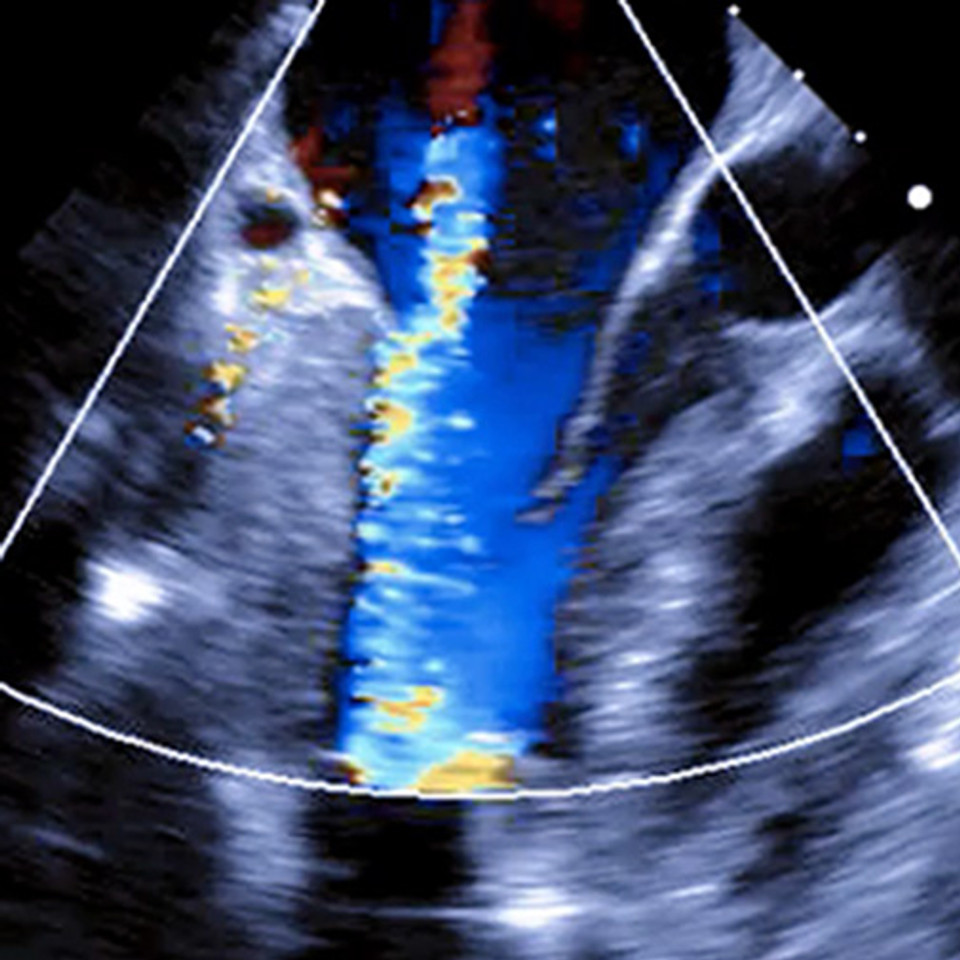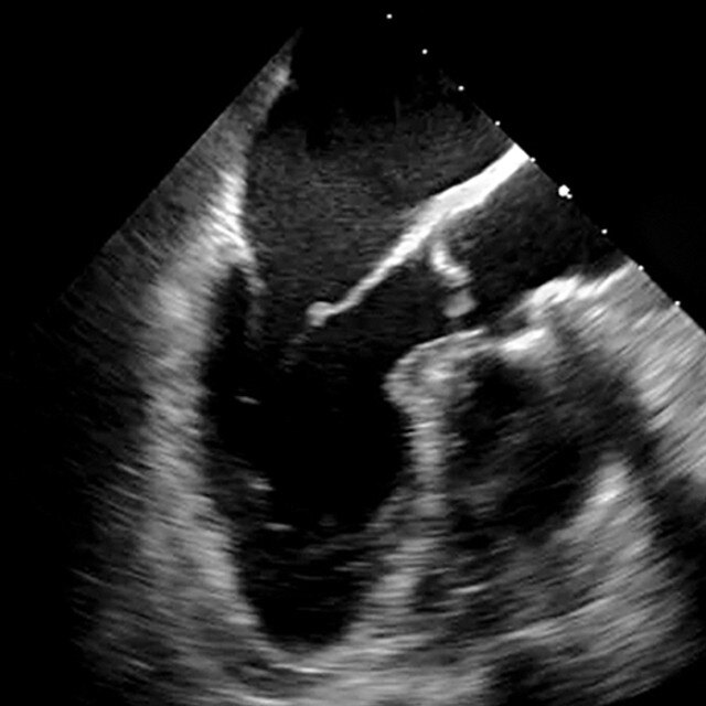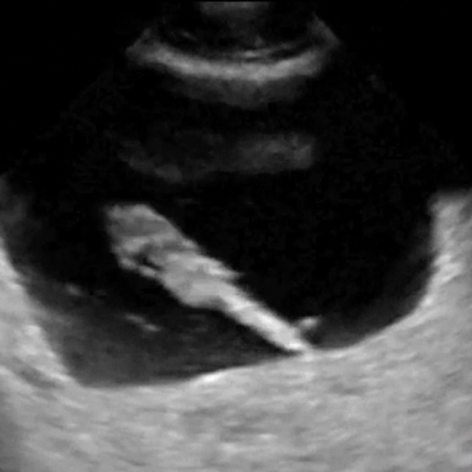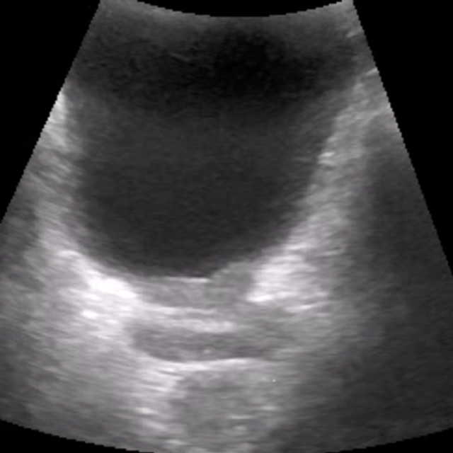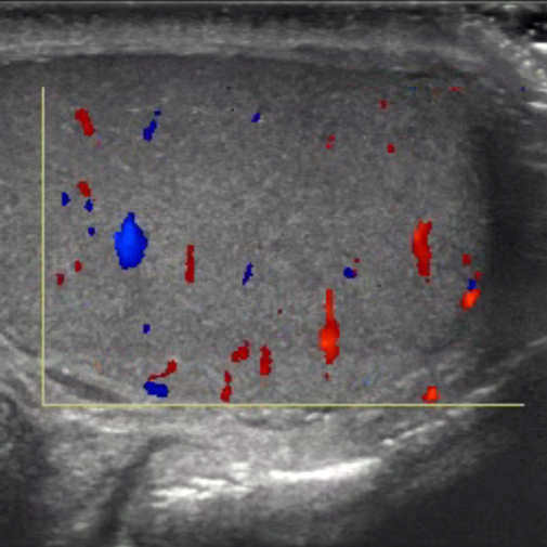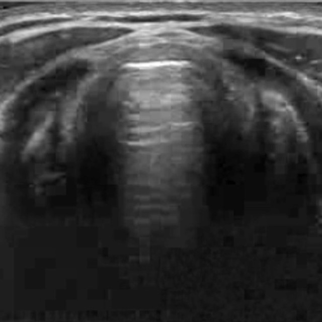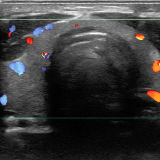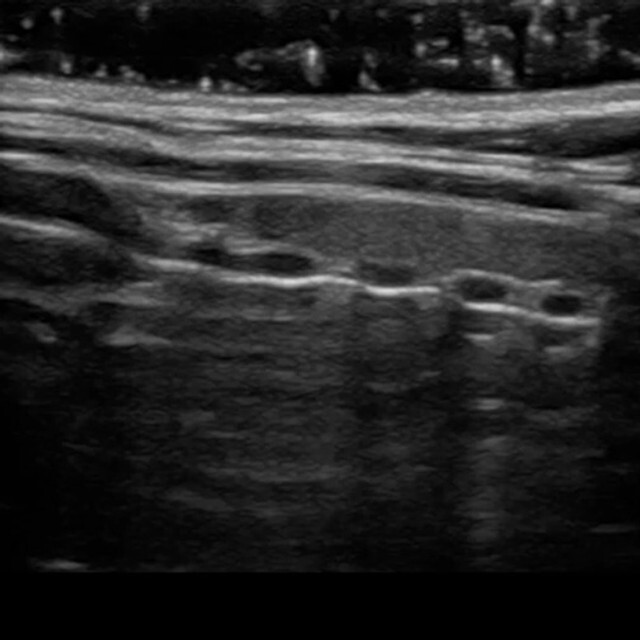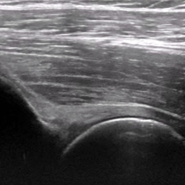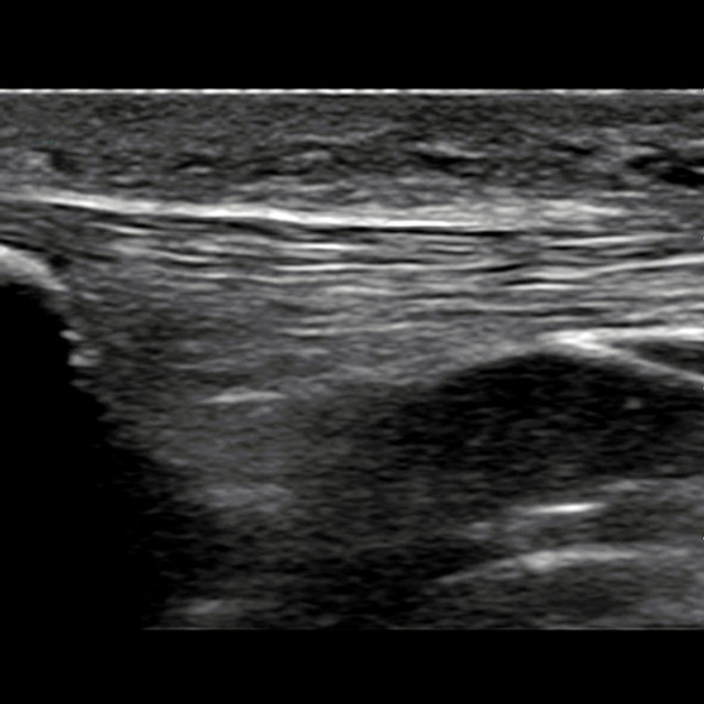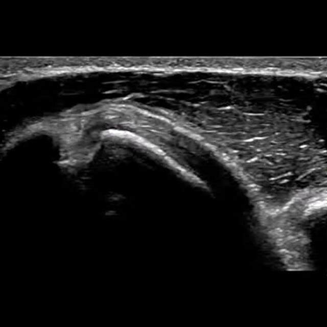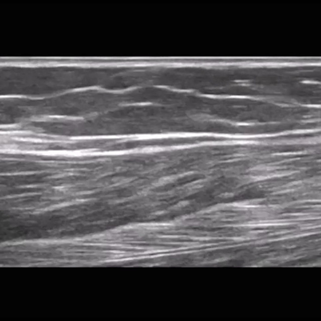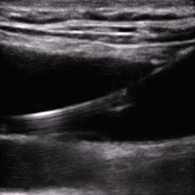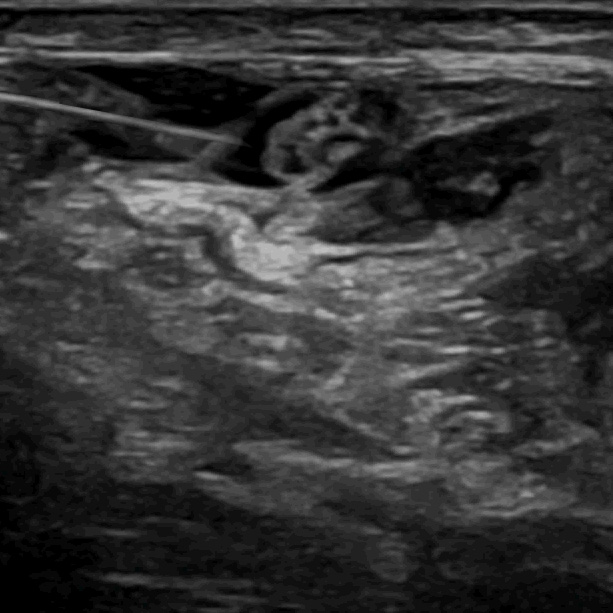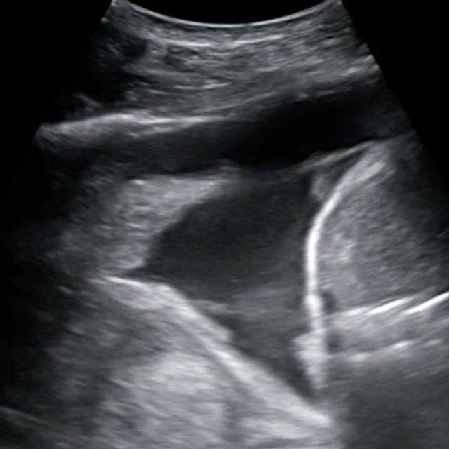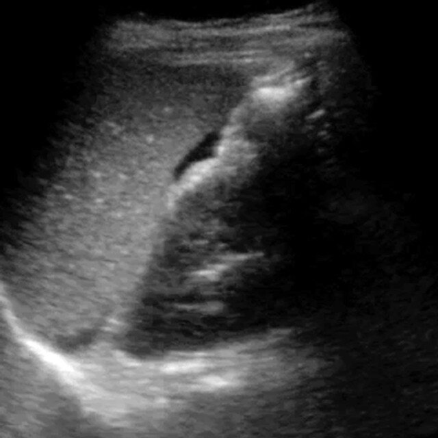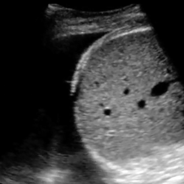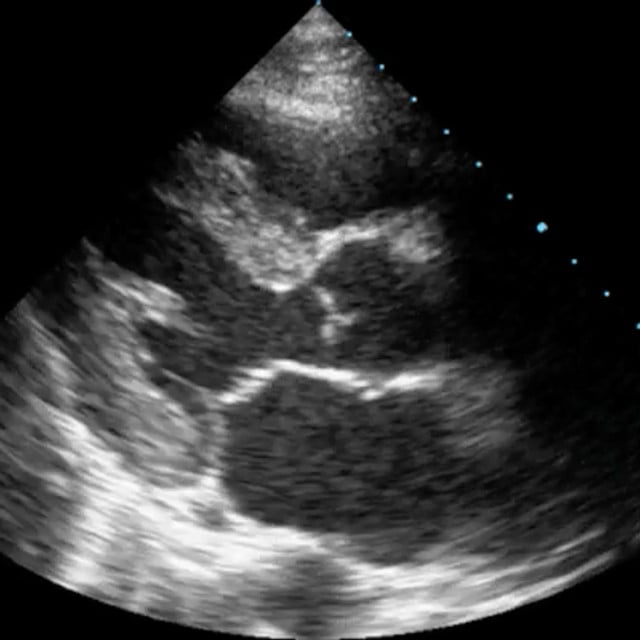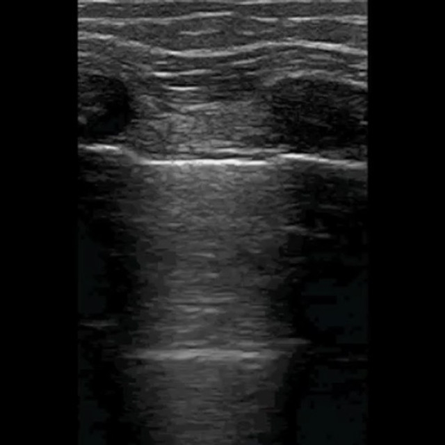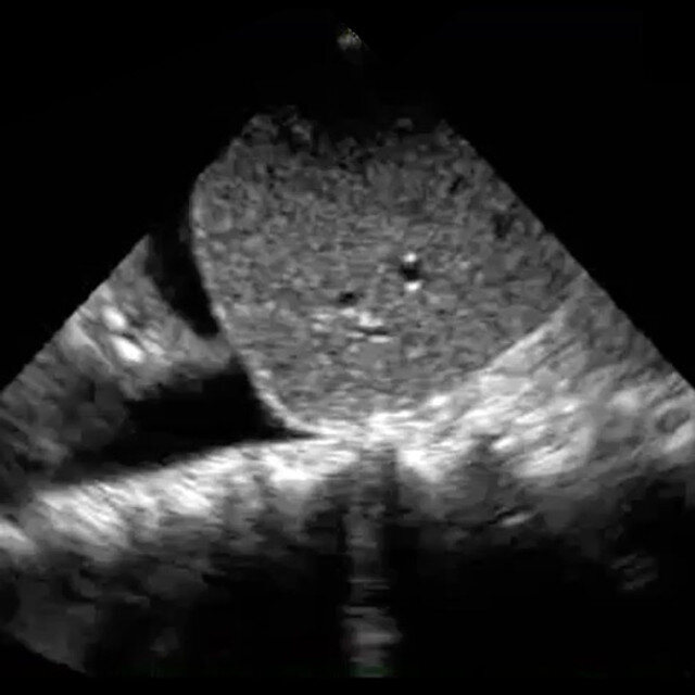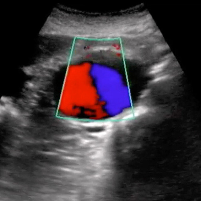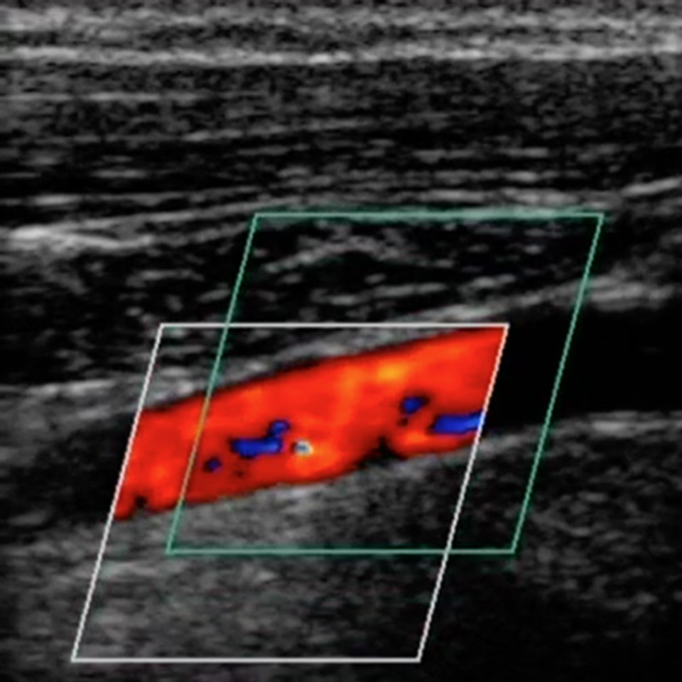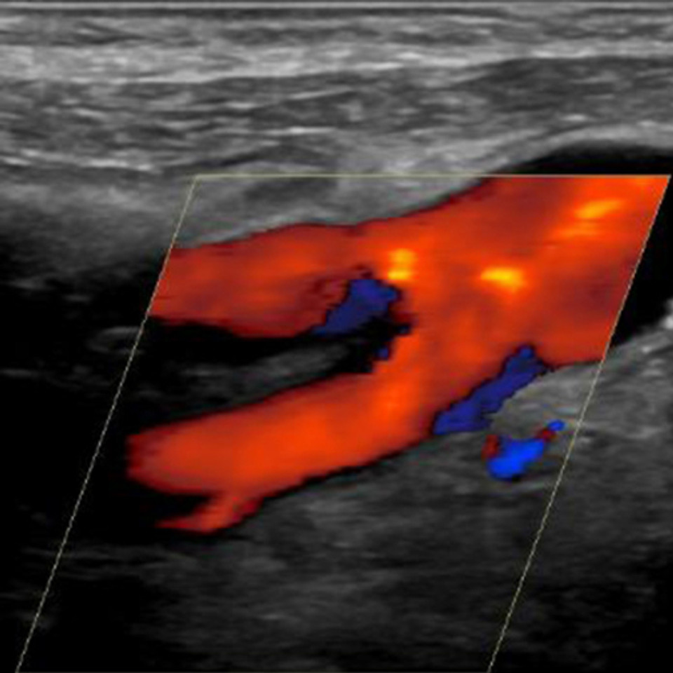
An Ultrasound Education Platform to Support Radiology Residency Programs

Standardize resident education in image acquisition and interpretation, expand access to hands-on practice with pathology, and track progress toward ultrasound proficiency.

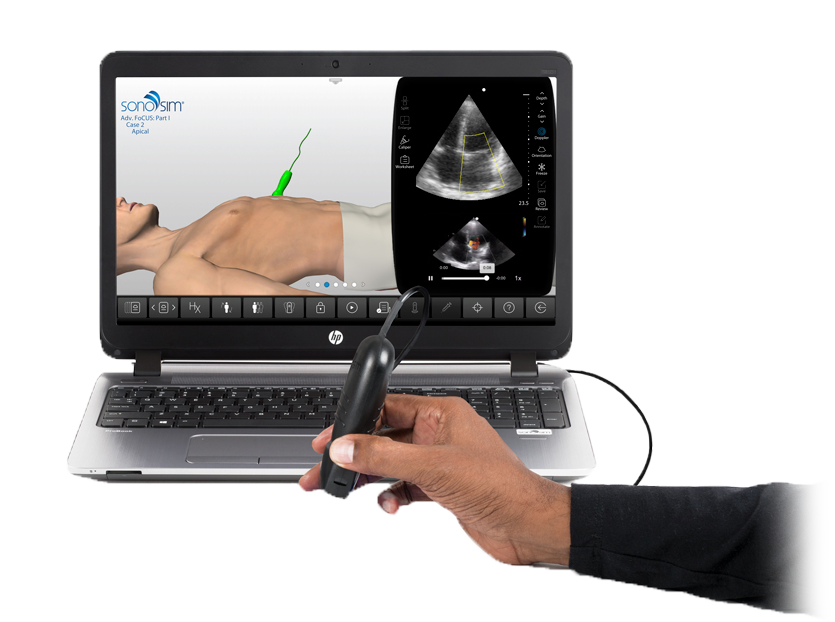
Simulator_image
clinical_dvt_product (1)_nsp

Build Ultrasound Proficiency Across Radiology Residency Programs

Give every resident a consistent foundation before clinical scanning begins.

Radiology residents must demonstrate competence in acquiring and interpreting ultrasound images. The 2026 ACGME requirements call for 75 hands-on scans and require programs to measure and document resident proficiency. ACGME guidance also recommends interpreting 150 examinations before graduation.¹

Clinical exposure alone can be uneven. Exam volume, case mix, and opportunities for residents to hold the transducer vary by rotation. SonoSim helps programs provide every resident with structured education, repeated scanning practice, and consistent exposure to normal and pathologic findings before and throughout clinical training.

For Radiology programs, this means:

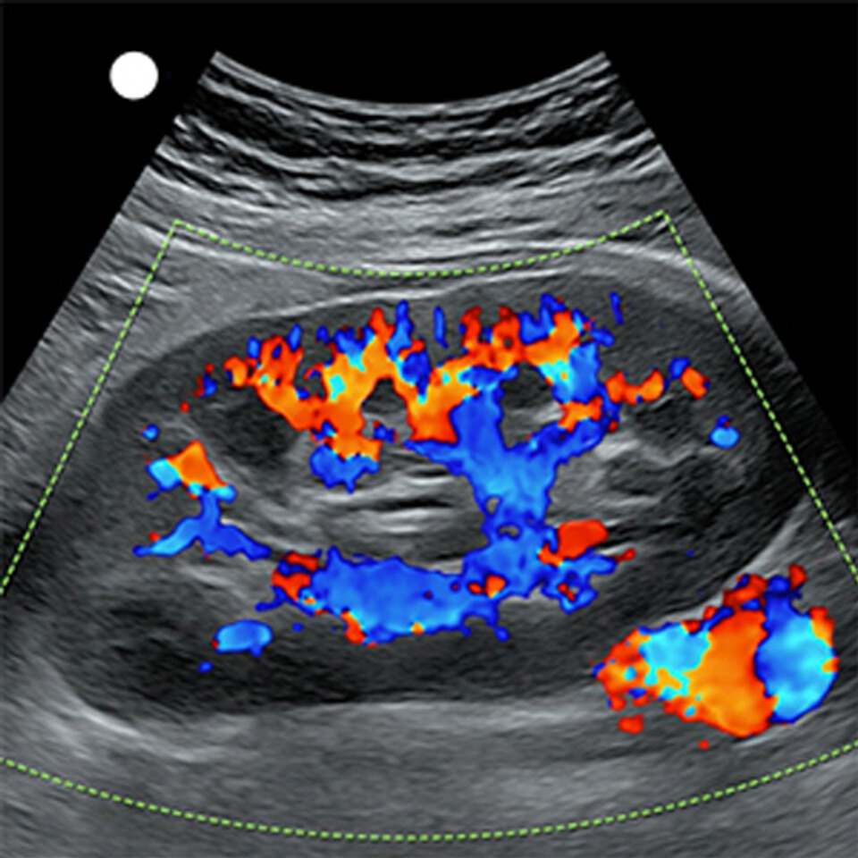
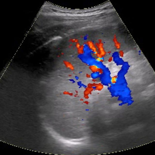
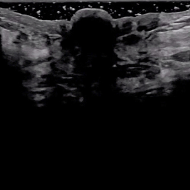
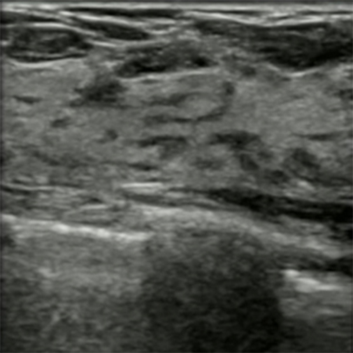
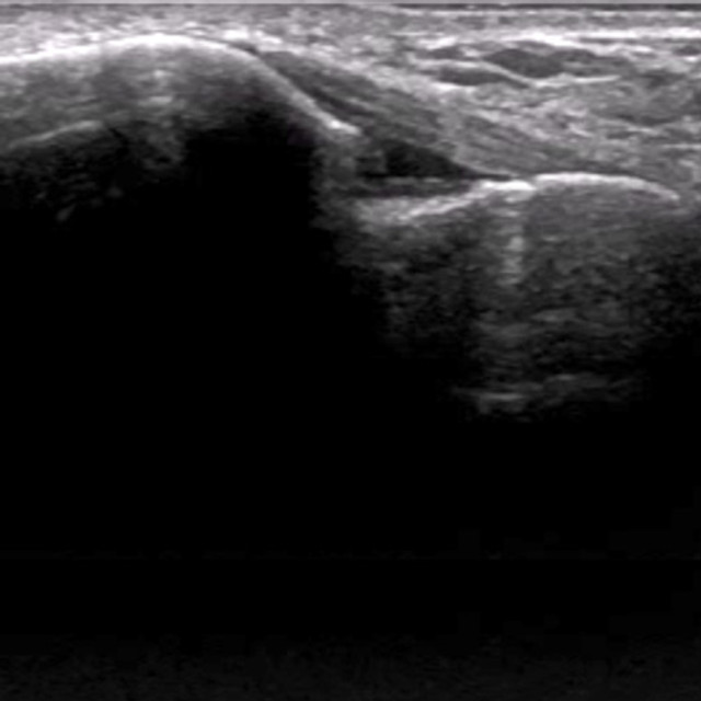
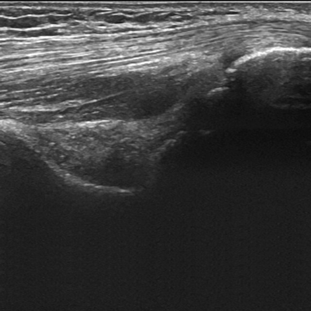
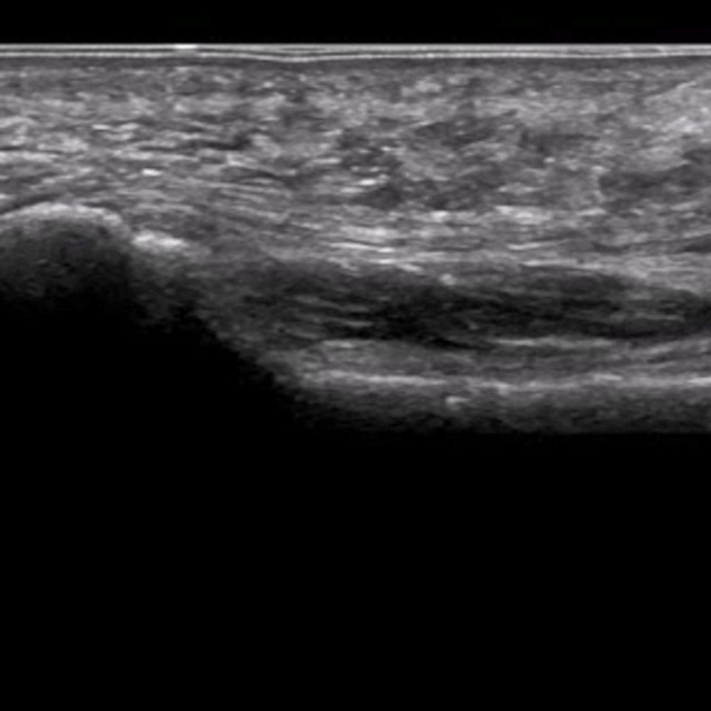
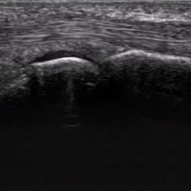
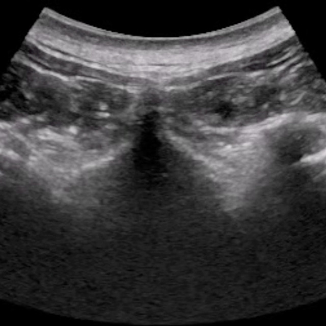
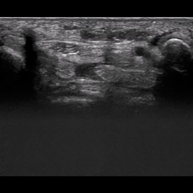
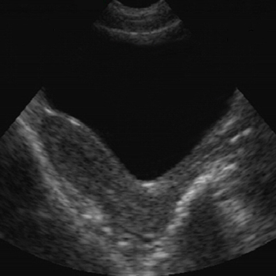
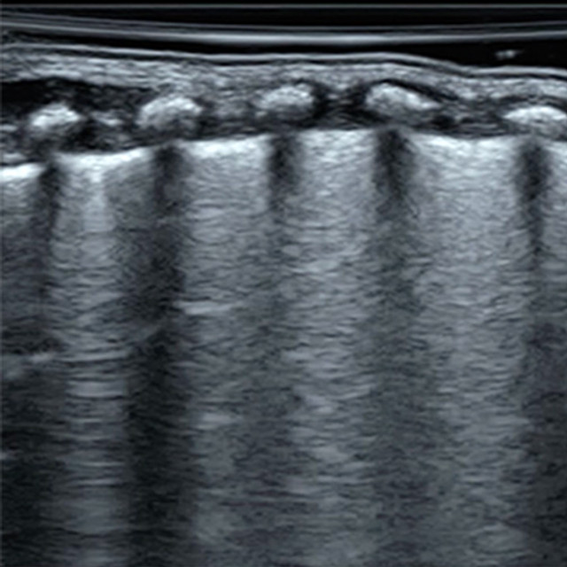
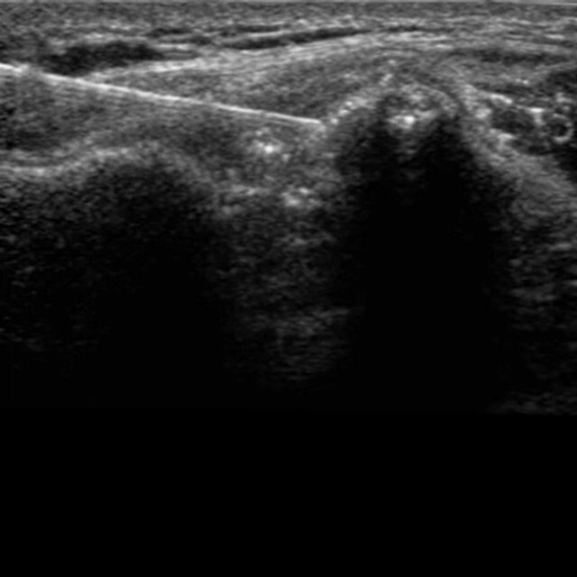
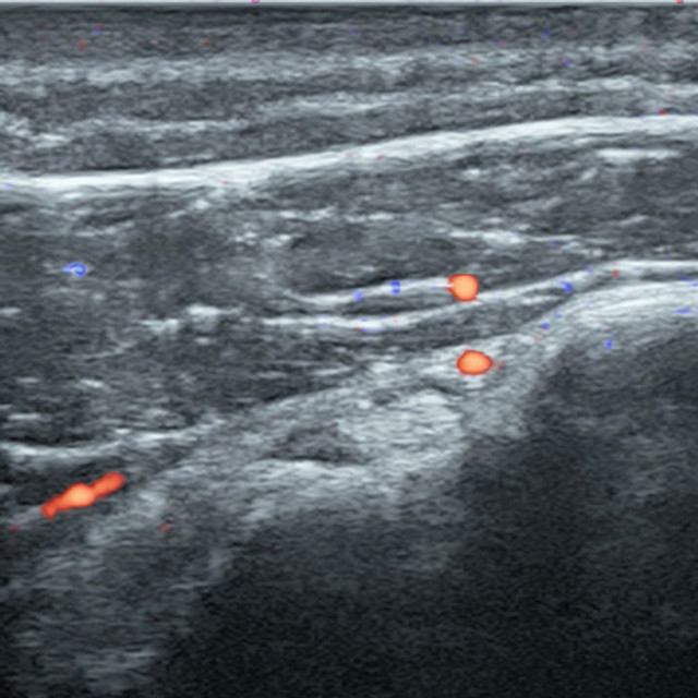
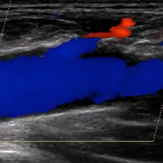
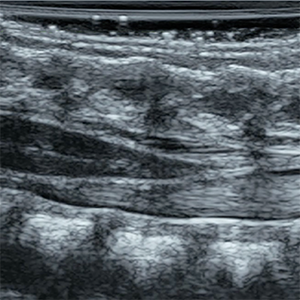
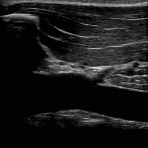
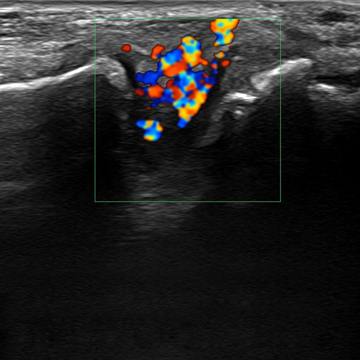
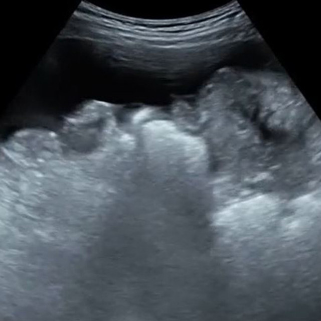
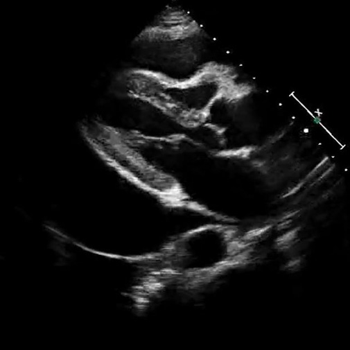
  • Building image acquisition and interpretation skills across abdominal, obstetric and gynecologic, vascular, pediatric, musculoskeletal, and breast applications
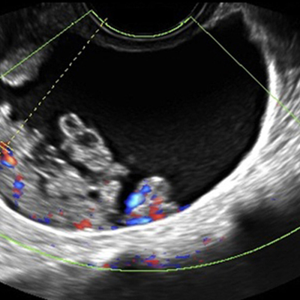
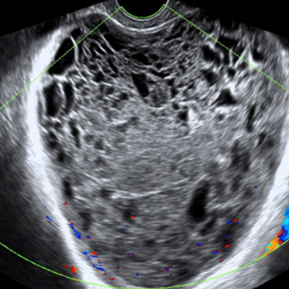
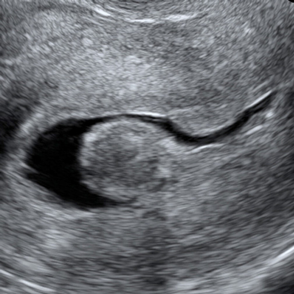
  • Providing repeatable practice with normal and pathologic cases beyond the variability of clinical rotations
  • Using consistent learning and performance data to monitor progress across residents and cohorts

A stronger foundation helps residents make better use of every supervised scanning opportunity.

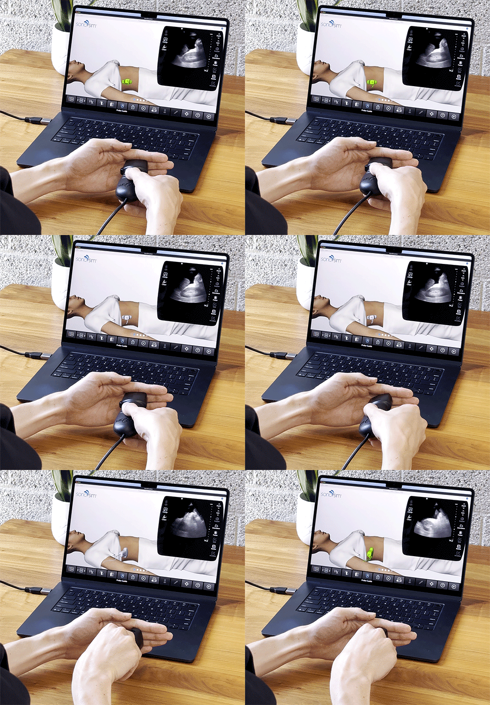
Product Image_2

Strengthen the Physics Foundation Behind Every Scan

Distributed review that supports the physics curriculum your program already leads.

Every ultrasound examination begins with an understanding of physics. The ACGME requires residents to demonstrate knowledge of medical imaging physics, including ultrasonography, and calls for clinical medical physics training throughout the 48 months of radiology education. The Review Committee also identifies ultrasound physics, knobology, image generation, and interpretation as foundational elements of sufficient hands-on experience.¹

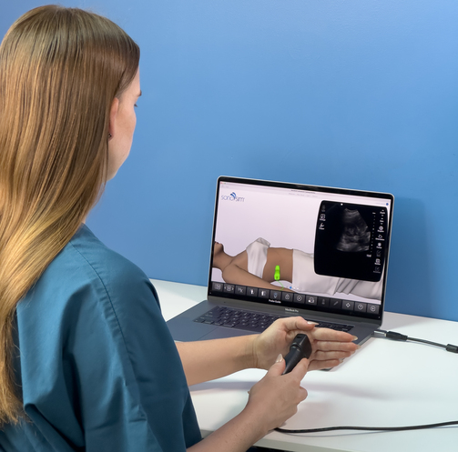
SonoSimulator Scanning_3.24.26.1

SonoSim gives residents a structured way to revisit these concepts on demand and connect them directly to scanning practice, complementing the program’s faculty-led and interactive physics education.

The SonoSim ecosystem includes:

  • Ultrasound physics review questions that residents can work through on their own schedule
  • Hands-on scanning practice in the SonoSimulator®, so residents build probe handling and image optimization before the rotation
  • Challenge Cases for quick, focused practice on advanced and uncommon pathology

Support Faculty and Scale Ultrasound Education

Tools to help Radiology educators teach, assess, and sustain a consistent curriculum.

The ACGME calls for designated physician faculty with expertise in and responsibility for developing ultrasonography didactics. It also requires the program’s overall core didactic curriculum to provide at least five hours of educational activity per week and repeat at least every two years.¹

SonoSim gives faculty a ready-to-deploy educational foundation, reducing the time required to rebuild ultrasound content while giving educators more opportunities to coach residents, review performance, and address individual learning needs.

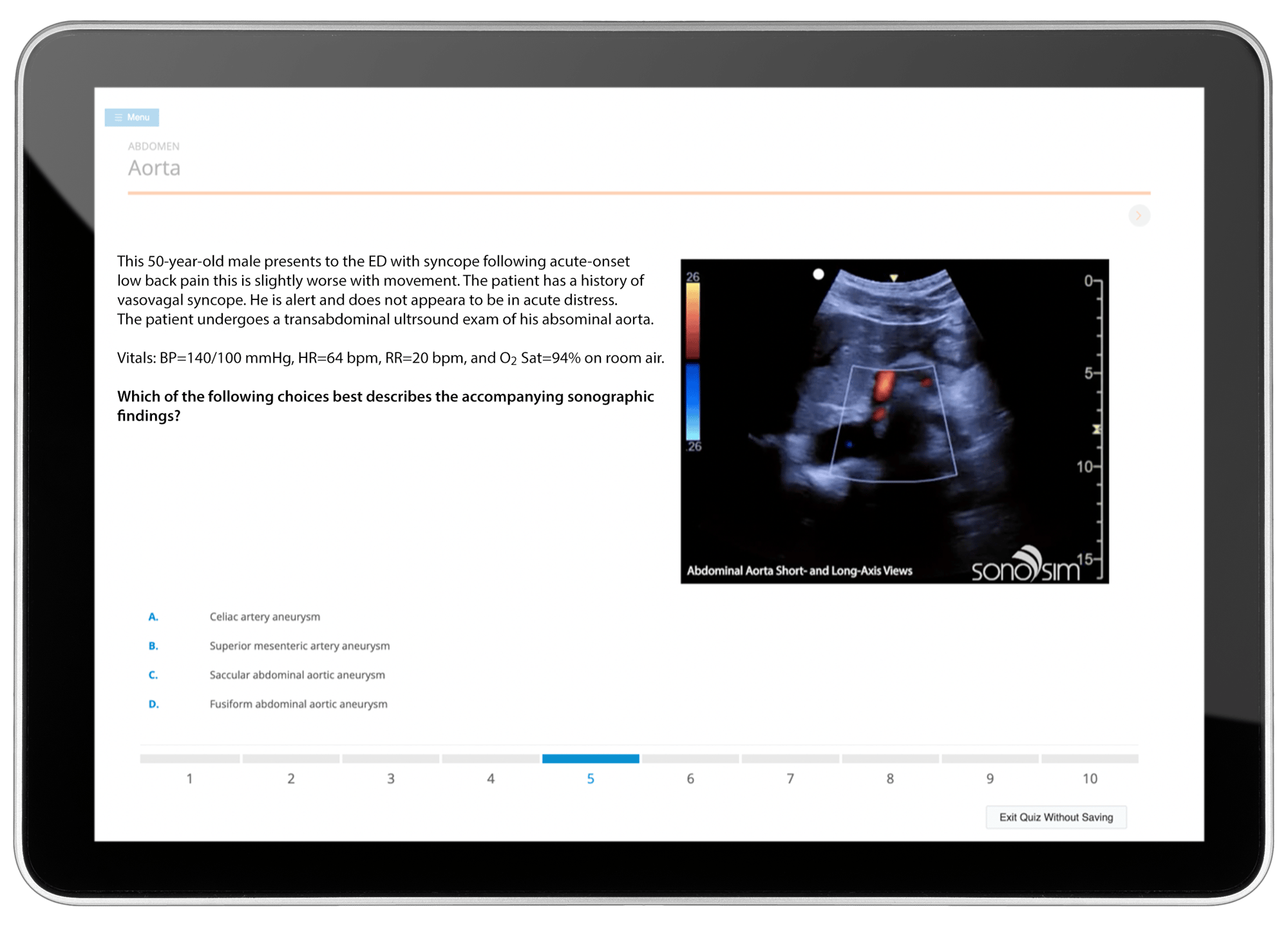
FPD_iPad_Final_090824.2

The SonoSim ecosystem supports Radiology programs with:

  • 85+ on-demand courses faculty can assign as the ultrasound didactic backbone, covering abdominal, OB/GYN, vascular, pediatric, MSK, and breast
  • A standardized case library so every resident in the cohort sees the same pathology, independent of what the rotation produces
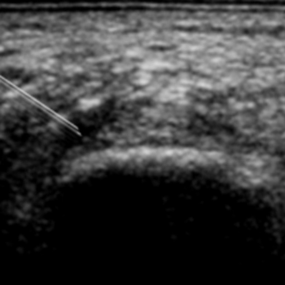
  • Ultrasound-guided procedure training across multiple applications
  • Performance tracking, image review, feedback tools, and learner-level portfolios
  • Curriculum integration support and customized curriculum mapping
  • A structured and proven† approach to building ultrasound competency
FAST_OTS_2

Ultrasound Education with SonoSim

Different Learners. Different Paths. One Trusted Platform. Ultrasound Education that Adapts to How You Teach.

Integrate Live Scanning into Radiology Training with SonoSim LiveScan®

Assess image acquisition and interpretation in realistic, standardized scenarios.

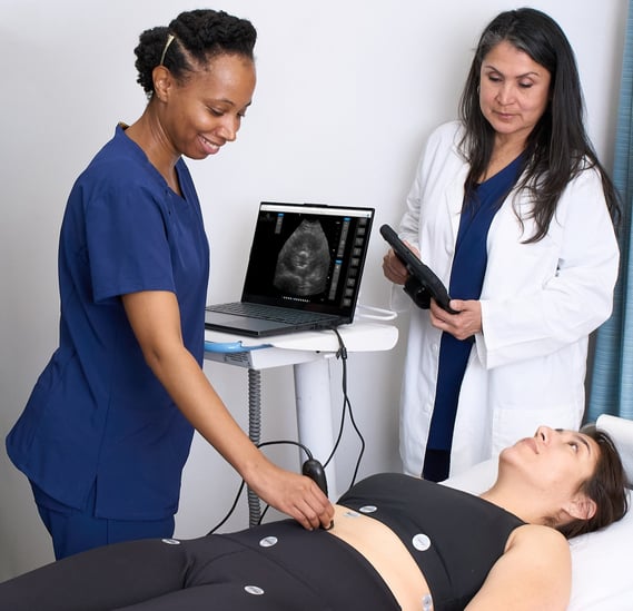
SonoSim LiveScan extends ultrasound education beyond the screen, allowing programs to evaluate how residents acquire and interpret ultrasound images in realistic, dynamic scenarios. By combining live scanning with simulated pathology, LiveScan helps faculty observe scanning technique, image acquisition, optimization, and interpretation as they happen.

2025_LiveScan_4-2
apply-3
  • Conduct repeatable scanning scenarios across Radiology applications
  • Instantly transform any manikin or live volunteer into a "patient" with real pathology
  • Run the same live scenario across a cohort, so faculty can compare performance on identical findings

Building Ultrasound Proficiency Across Radiology Programs

A structured, program-level approach to training, assessment, and documentation.

Proficiency depends on how well each resident scans and interprets, not just how many examinations they complete, and the ACGME expects programs to document it.¹  The ACR–SPR–SRU practice parameter describes diagnostic ultrasound as protocol-driven, with defined standardized views and documented images and interpretation. It also recognizes completion of an accredited radiology residency as one pathway to being qualified to perform, interpret, and report those examinations.²  What a program builds during residency is what its graduates carry into practice.

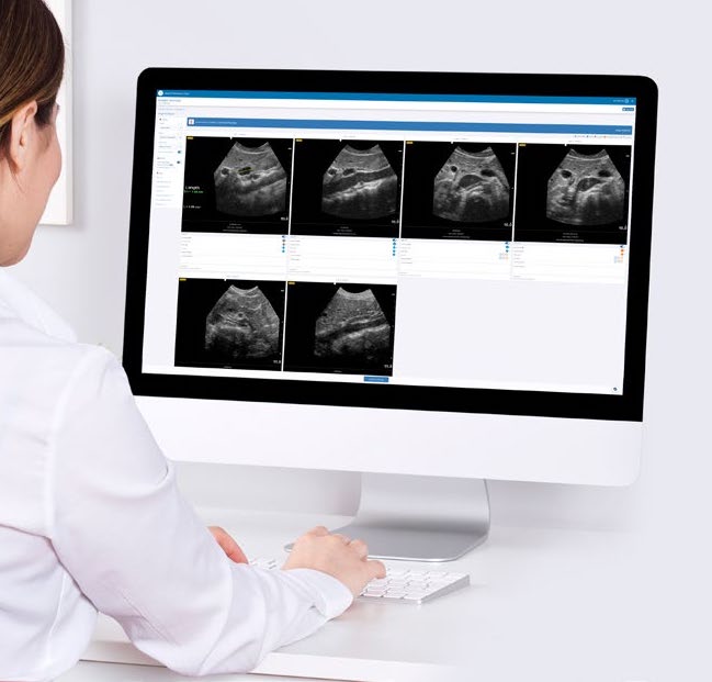
SonoSim Performance Tracker gives faculty a structured way to see that proficiency develop. Scanning work in the SonoSimulator, course progress, and QuestionBank activity are recorded for each resident, and faculty can review captured images and give feedback directly. Each resident's work builds into an ultrasound e-portfolio that programs can reference alongside their own evaluation process.

Because every resident's record is built on the same case set, it is comparable across the cohort, giving program leadership a consistent view of ultrasound skill as residents progress through the program.

AIA-1

Covering Ultrasound Applications Across Radiology Requirements

Organized around the diagnostic ultrasound applications, Radiology residents train in

SonoSim supports Radiology ultrasound education across the diagnostic applications residents perform and interpret during training. Content is structured to support standardized, competency-based training, with curricular mapping to the ACGME Diagnostic Radiology Program Requirements and alignment with the ACR–SPR–SRU practice parameter for diagnostic ultrasound. Customized curriculum mapping is also available on a program-by-program basis for SonoSim clients.


Mapped to the ACGME Diagnostic Radiology Program Requirements

Physics

Ultrasound Physics & Fundamentals
 

Abdomen

Adrenal Glands
Adrenal Glands
 
 
Intestinal & Biliary Clinical
 

Breast

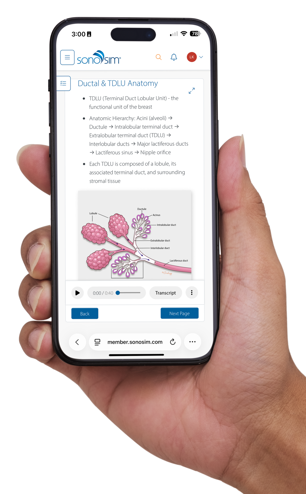
Introduction to Breast Ultrasound
 

Cardiac

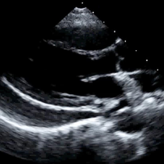
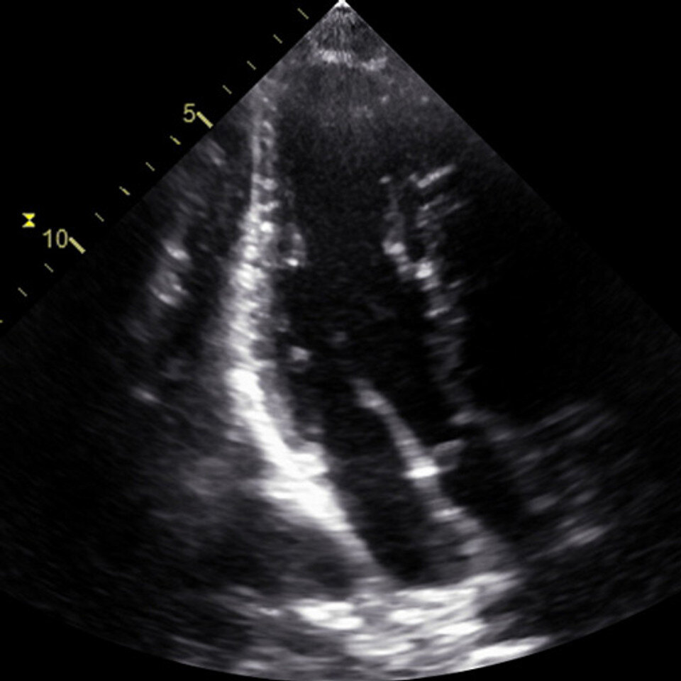
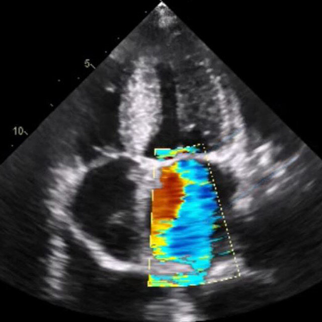
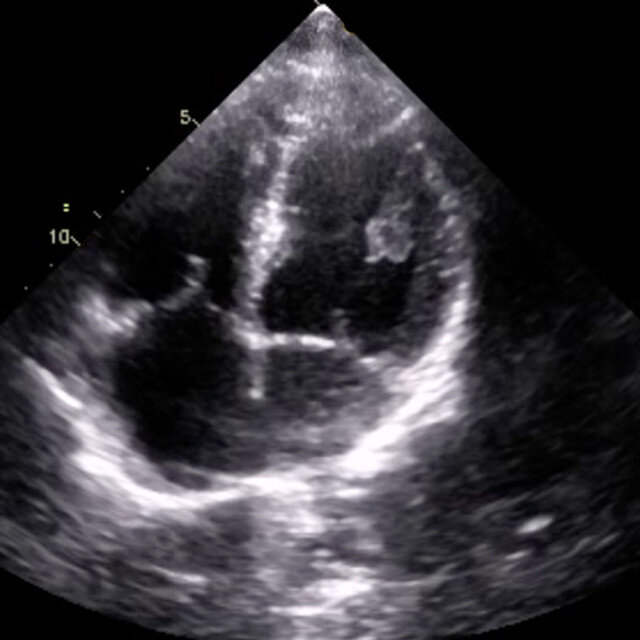
Cardiology Clinical
Cardiology Clinical
 
FoCUS Advanced Clinical Part 1
 
FoCUS Advanced Clinical Part 2
 
LVAD Problem-Focused Echocardiography
 
Resuscitative Transesophageal Echocardiography (TEE)
 

Genitourinary

Head/Neck

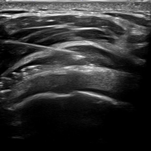
Musculoskeletal

Obstetrics/Gynecology 

Basic Obstetric – Six-Step Approach
 
Female Pelvis
Female Pelvis
 
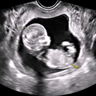
First-Trimester Pregnancy
 
 
Second and Third Trimester Pregnancy Part 1
 
Second and Third Trimester Pregnancy Part 2
 
Ultrasound Malignant Adnexal Conditions
 
Ultrasound Nonmalignant Adnexal Conditions
 
Ultrasound of Nonpregnant Abnormal Uterus Part 1
 
Ultrasound of Nonpregnant Abnormal Uterus Part 2
 
Ultrasound of Nonpregnant Normal Adnexa
 
Ultrasound of Nonpregnant Normal Uterus
 

Ocular


Pediatric

Neonatal & Infant Neurosonography
 
Pediatric Lung Sonography
 

Procedures

Acromioclavicular Joint Injection & Aspiration
 
Biceps Tendon Sheath Injection
 
Femoral Line Placement
 
Glenohumeral Joint Injection
 
Internal Jugular Vein Cannulation
 
Knee Joint Injection & Aspiration
 
Metatarsophalangeal Joint Injection & Aspiration
 
Neonatal & Infant Lumbar Puncture
 
Peripheral Venous Access
 
Plantar Fasciitis Ultrasound-Guided Injection
 
Subacromial-Subdeltoid Bursa Injection & Aspiration
 
Subclavian Vein Cannulation
 
Tibiotalar Joint Injection & Aspiration
 
Ultrasound-Guided Paracentesis Procedure
 
Ultrasound-Guided Pericardiocentesis Procedure
 
Ultrasound-Guided Procedures Introductory
 
Ultrasound-Guided Regional Anesthesia
 
Ultrasound-Guided Thoracentesis Procedure
 

Protocols

eFAST Protocol
eFAST Protocol
 
FAST Protocol
FAST Protocol
 
RUSH Protocol
RUSH Protocol
 

Pulmonary

Pulmonary Clinical
Pulmonary Clinical
 

Vascular

Aorta & Inferior Vena Cava (IVC)


Cerebrovascular
Cerebrovascular
 
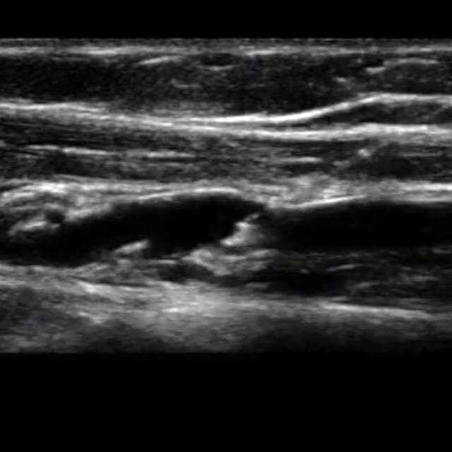
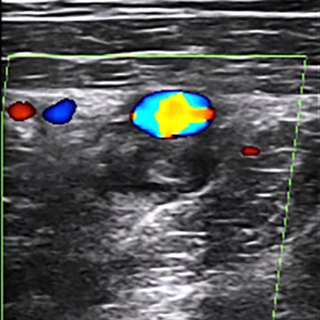
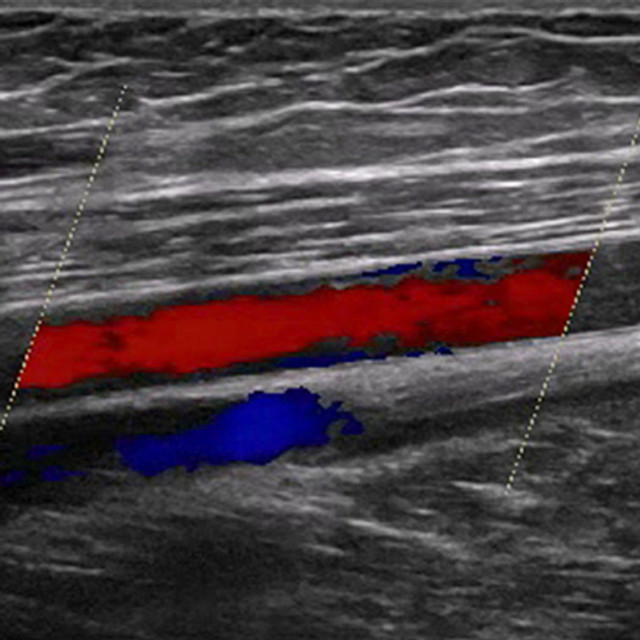
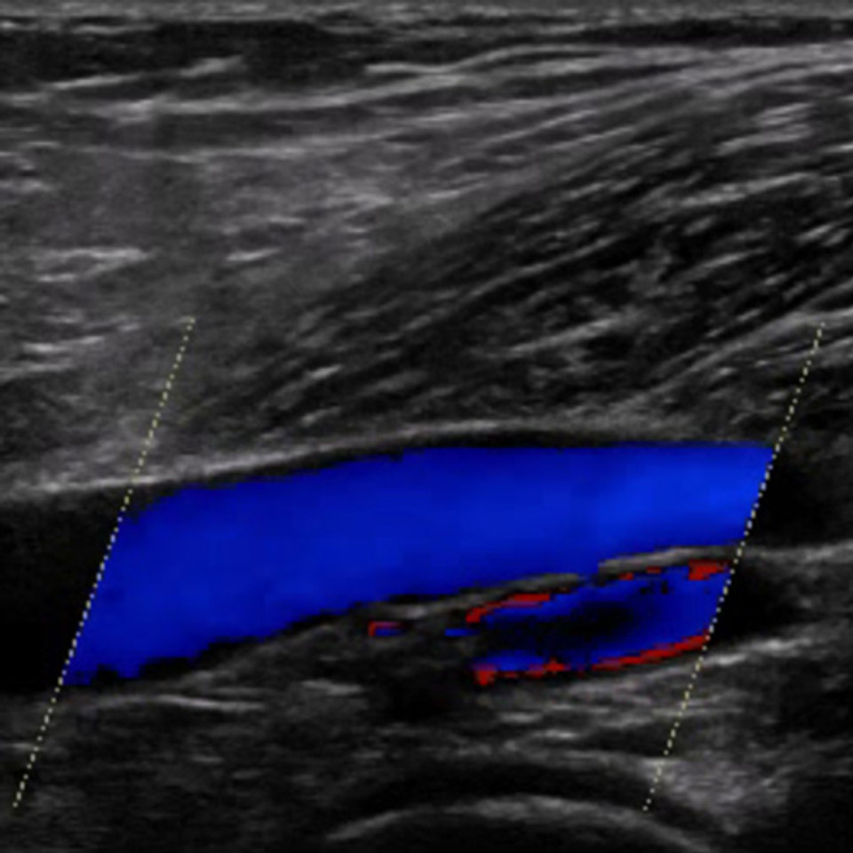
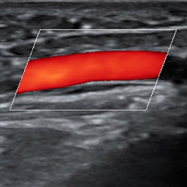
Deep Vein Thrombosis (DVT) Lower Extremity


Vascular Mapping for Preoperative Planning of Dialysis Access
 
Intro_to_breast_phone_mock_up

In one platform, Radiology programs get:

  • Structured didactic learning: On-demand ultrasound education covering anatomy & physiology, as well as core, advanced, and procedural applications relevant to Radiology.
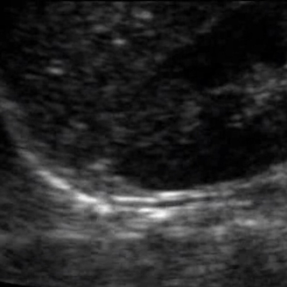
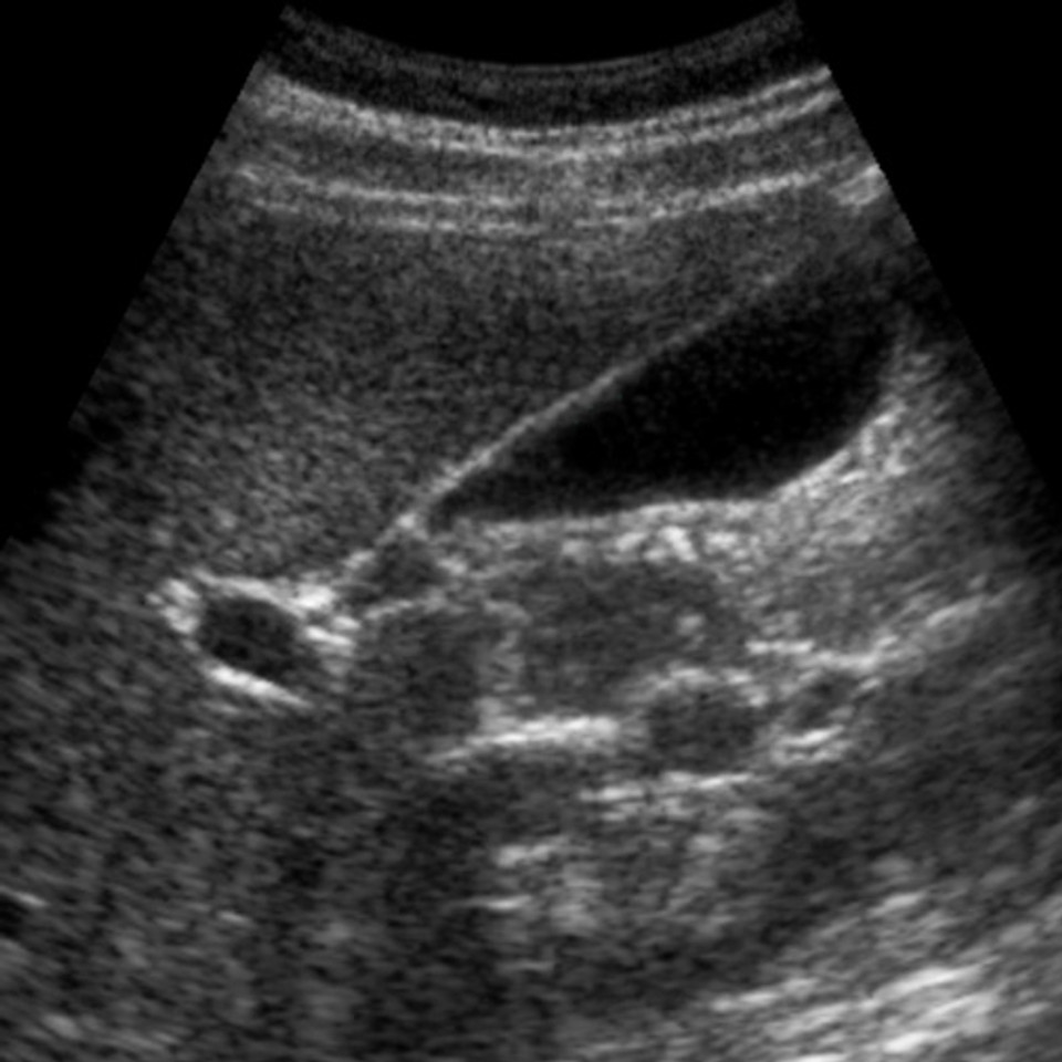
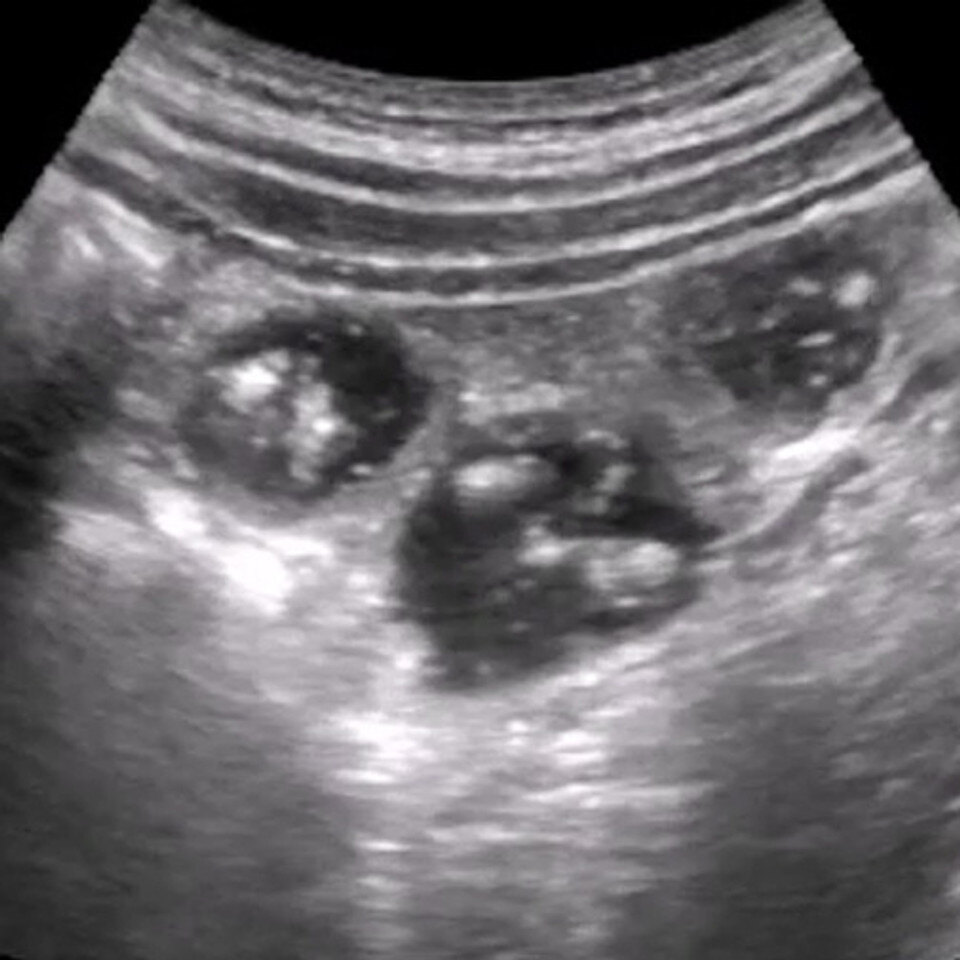
  • SonoSimulator® scanning with pathology: Anytime, anywhere access to scanning cases from real patients that reinforce image acquisition, optimization, interpretation, and pattern recognition
  • Deliberate practice and challenge cases: Opportunities for learners to apply ultrasound findings in clinically meaningful scenarios that mirror real-world diagnostic practice.
  • Performance assessment and progress tracking: Tools that help faculty monitor learner development, support consistent assessment, and identify gaps across cohorts.
  • Flexible integration into existing curricula: A scalable platform designed to complement existing Radiology curricula without adding unnecessary administrative burden

Review the full SonoSim Ecosystem

  • Learn

  • Scan

  • Practice

  • Track

  • Study

  • Apply

  • Integrate

Learn

Interactive Ultrasound Courses

Explore SonoSim's comprehensive library of on-demand online ultrasound courses covering thousands of ultrasound applications. Authored by leading experts, our interactive and multimedia courses combine regional anatomy, sonographic insight, practical imaging techniques, real patient cases, and helpful videos. Get engaged with our CME*-eligible blend of learning tools.
Scan

Hands-On Virtual Ultrasound Scanning

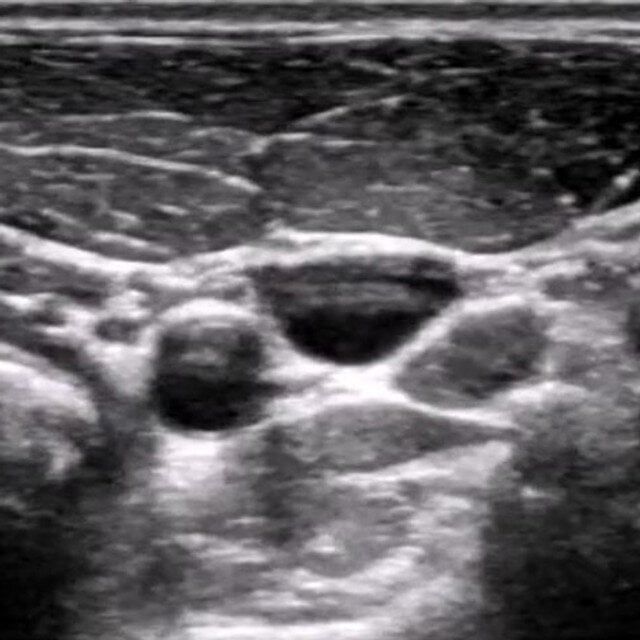
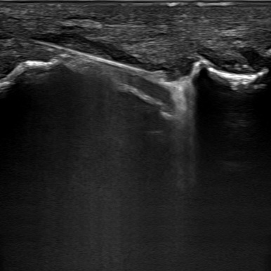
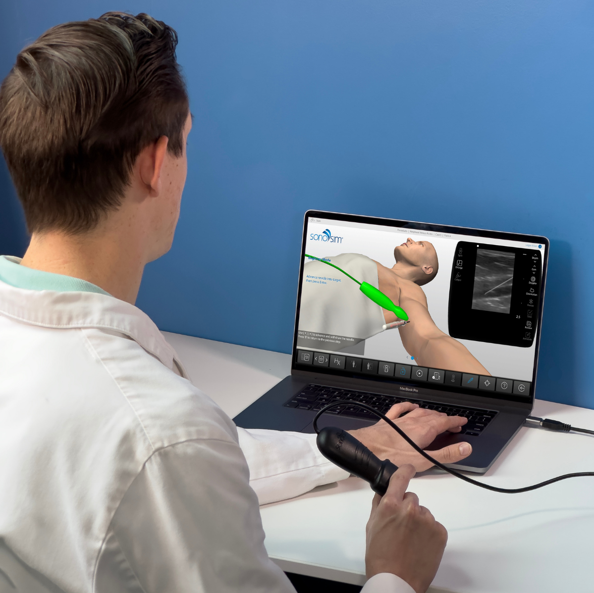
Unlock hands-on ultrasound experience with the SonoSimulator®. This proven methodology breaks down the traditional barriers to ultrasound learning, offering access to thousands of real patient scanning opportunities of pathologic conditions.

For beginners and practicing physicians alike, our software provides on-demand, AI-driven performance assessments, guided probe positioning, and immediate feedback. Learn to acquire and interpret ultrasound images on a laptop, with support from a virtual tutor, right at your fingertips.
practice

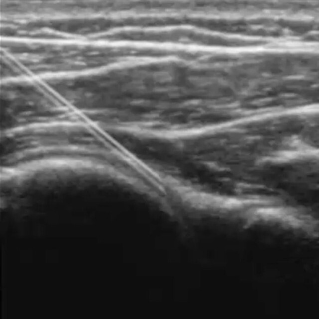
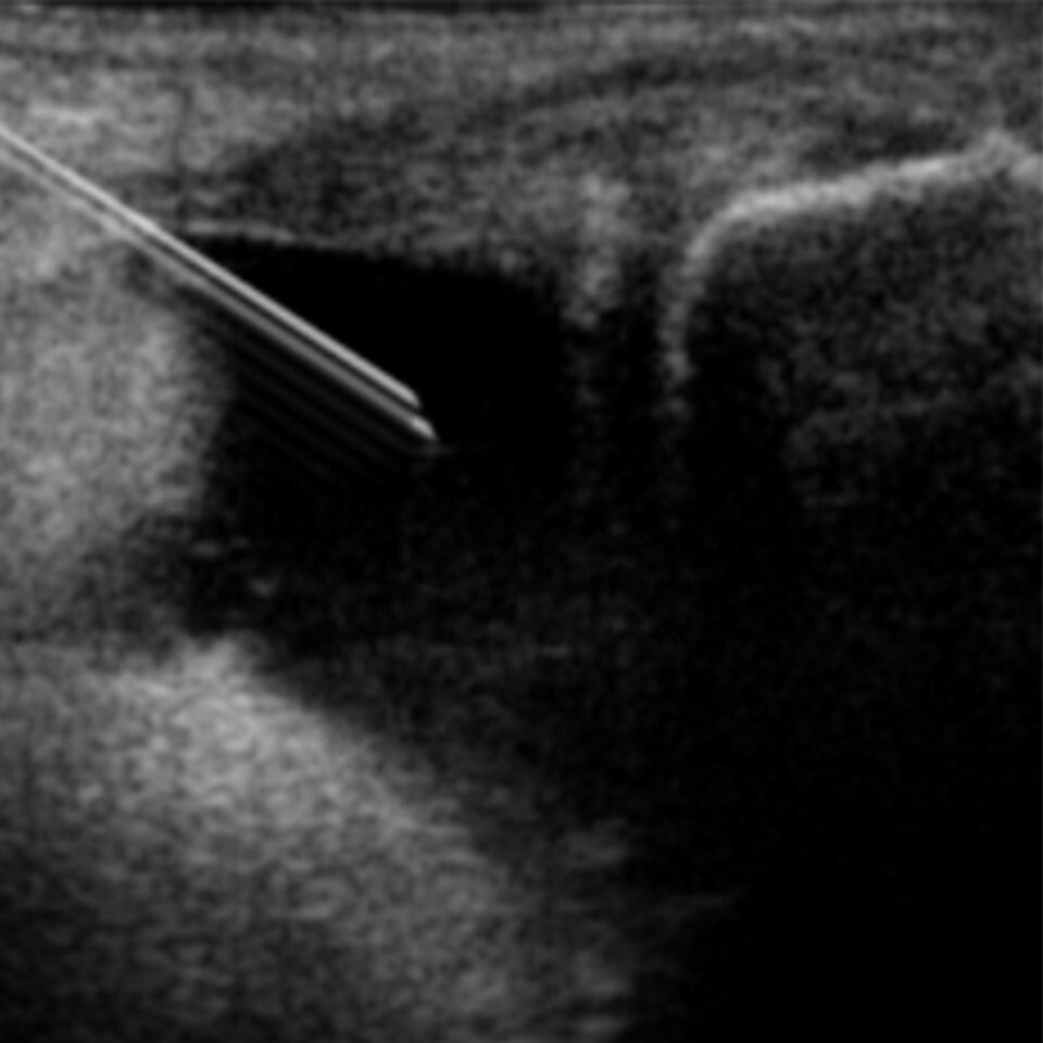
Risk-Free Practice of Ultrasound-Guided Procedures

Real-time ultrasound guidance for procedures like peripheral IV access is rapidly becoming the standard of care, demanding not only significant knowledge but also refined psychomotor skills.

SonoSim's training environment offers learners a safe and convenient space to hone these essential abilities. With unrestricted access to deliberate practice, SonoSim users can build the knowledge, skill, and confidence needed for success in ultrasound-guided procedures.
track-1

Track & Assess Progress

SonoSim revolutionizes learner assessment by offering comprehensive tools to monitor progress, deliver feedback, and even automate ultrasound image review. These features combine to alleviate the most time-consuming parts of managing ultrasound programs, allowing for more focused and effective training. And for learners, this feature provides access to ultrasound image portfolios which are useful for job and internship applications. 
study-3

Build & Hone Knowledge with Study Tools & Resources

Now, more accessible than ever, learners can review and study ultrasound on-the-go. With case-based learning in our SonoSim QuestionBank, ultrasound case studies become powerful tools to test and reinforce knowledge. Plus, we offer an SPI exam study guide and ultrasound physics review questions, catering to learners of all backgrounds and needs. Our Challenge Cases give learners quick cases to challenge their skills. And a mobile app provides key references on any device.
apply-3

Bring Ultrasound Simulation to Life

SonoSim LiveScan® offers an immersive, versatile tool for teaching & evaluating the integration of ultrasound into medical decision-making. Whether used with live volunteers or existing manikins, SonoSim LiveScan seamlessly incorporates ultrasound into scenario-based medical simulations, providing more realistic and enriching learning experiences.
integrate-2

Integrate SonoSim into Your Curriculum

We work closely with you to construct a thriving ultrasound training program tailored to your organization's needs. SonoSim's all-encompassing approach, featuring tools, expert guidance, resources, and support, ensures a smooth incorporation of ultrasound education into your existing curriculum. By empowering both educators and learners, our ecosystem unlocks the full potential of ultrasound, providing a pathway to success.

puzzle strip2

Let’s Talk About Ultrasound Education in Your Radiology Residency Program

Learn how SonoSim can help standardize ultrasound training, expand access to pathology, and track resident progress toward proficiency.